Application
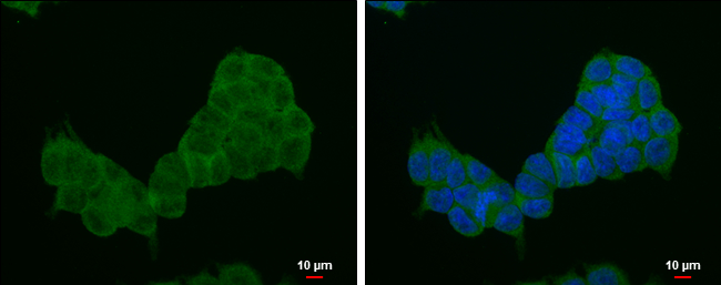
4E-BP1 antibody [N1C3-2] detects 4E-BP1 protein at cytoplasm by immunofluorescent analysis. Sample: HCT116 cells were fixed in 4% paraformaldehyde at RT for 15 min. Green: 4E-BP1 protein stained by 4E-BP1 antibody [N1C3-2] diluted at 1:1000. Blue: Hoechst 33342 staining. Scale bar = 10 μm.Application
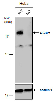
Wild-type (WT) and 4E-BP1 knockout (KO) HeLa cell extracts (30 µg) were separated by 15% SDS-PAGE, and the membrane was blotted with 4E-BP1 antibody [N1C3-2] diluted at 1:1000. The HRP-conjugated anti-rabbit IgG antibody was used to detect the primary antibody, and the signal was developed with Trident ECL plus-Enhanced.Application
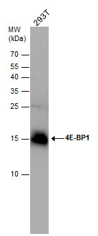
4E-BP1 antibody detects 4E-BP1 protein by western blot analysis. Various whole cell extracts (30 μg) were separated by 15% SDS-PAGE, and the membrane was blotted with 4E-BP1 antibody diluted at a dilution of 1:1000.| Product Name | 4E-BP1 antibody |
|---|---|
| Antibody Type | Primary Antibodies |
| Product description | Rabbit Polyclonal antibody to 4E-BP1 (eukaryotic translation initiation factor 4E binding protein 1)This gene encodes one member of a family of translation repressor proteins. The protein directly interacts with eukaryotic translation initiation factor 4E (eIF4E), which is a limiting component of the multisubunit complex that recruits 40S ribosomal subunits to the 5' end of mRNAs. Interaction of this protein with eIF4E inhibits complex assembly and represses translation. This protein is phosphorylated in response to various signals including UV irradiation and insulin signaling, resulting in its dissociation from eIF4E and activation of mRNA translation. [provided by RefSeq] |
| Immunogen | Full length human 4E-BP1 Recombinant protein. |
| Clonality | Polyclonal |
|---|---|
| Isotype | IgG |
| Host Species | Rabbit |
| Tested Applications | ICC/IFWB |
| *Optimal dilutions/concentrations should be determined by the researcher.
: |
|
| Species Reactivity | Human |
| Predicted Reactivity | Mouse, Rat, Pig, BovineMouse (90%), Rat (92%), Pig (91%), Bovine (92%) |
| Concentration | 1mg/ml |
| Purification | Affinity purified |
| Gene Synonyms | 4E-BP1 antibody 4EBP1 antibody BP-1 antibody MGC4316 antibody PHAS-I antibody EIF4EBP1 antibody eukaryotic translation initiation factor 4E-binding protein 1 antibody eIF4E-binding protein 1 antibody phosphorylated heat- and acid-stable protein reg |
|---|---|
| Molecular Weight(MW) | 13 kDa (note) (2) |
Application
4E-BP1 antibody [N1C3-2] detects 4E-BP1 protein at cytoplasm by immunofluorescent analysis. Sample: HCT116 cells were fixed in 4% paraformaldehyde at RT for 15 min. Green: 4E-BP1 protein stained by 4E-BP1 antibody [N1C3-2] diluted at 1:1000. Blue: Hoechst 33342 staining. Scale bar = 10 μm.
Application
Wild-type (WT) and 4E-BP1 knockout (KO) HeLa cell extracts (30 µg) were separated by 15% SDS-PAGE, and the membrane was blotted with 4E-BP1 antibody [N1C3-2] diluted at 1:1000. The HRP-conjugated anti-rabbit IgG antibody was used to detect the primary antibody, and the signal was developed with Trident ECL plus-Enhanced.
Application
4E-BP1 antibody detects 4E-BP1 protein by western blot analysis. Various whole cell extracts (30 μg) were separated by 15% SDS-PAGE, and the membrane was blotted with 4E-BP1 antibody diluted at a dilution of 1:1000.| Positive Control | 293T , HeLa |
|---|---|
| Application Notes | *Optimal dilutions/concentrations should be determined by the researcher.
: |
| Form | Liquid |
|---|---|
| Storage Instructions | Keep as concentrated solution. Aliquot and store at -20ºC or below. Avoid multiple freeze-thaw cycles. |
| Storage Buffer | 1XPBS, 20% Glycerol (pH7). 0.025% ProClin 300 was added as a preservative. |


