Application
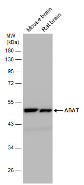
Various tissue extracts (50 µg) were separated by 10% SDS-PAGE, and the membrane was blotted with ABAT antibody diluted at 1:10000. The HRP-conjugated anti-rabbit IgG antibody was used to detect the primary antibody.Application
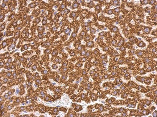
ABAT antibody detects ABAT protein at mitochondria on mouse liver by immunohistochemical analysis. Sample: Paraffin-embedded mouse liver. ABAT antibody dilution: 1:500.Application
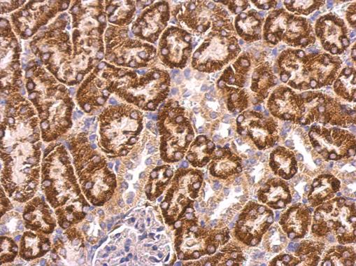
ABAT antibody detects ABAT protein at mitochondria on mouse kidney by immunohistochemical analysis. Sample: Paraffin-embedded mouse kidney. ABAT antibody dilution: 1:500.Application
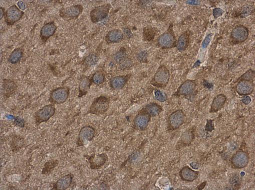
ABAT antibody detects ABAT protein at cytoplasm in rat brain by immunohistochemical analysis. Sample: Paraffin-embedded rat brain. ABAT antibody diluted at 1:500.Application
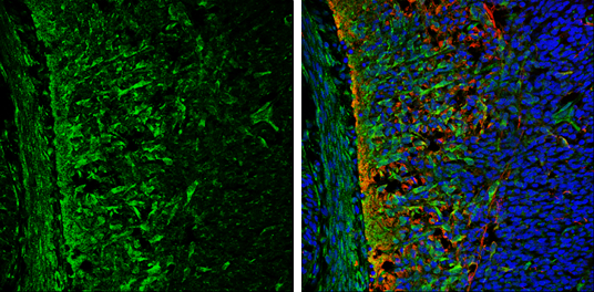
ABAT antibody detects ABAT protein expression by immunohistochemical analysis. Sample: Frozen sectioned E13.5 Rat brain. Green: ABAT protein stained by ABAT antibody diluted at 1:250. Red: beta Tubulin 3/ TUJ1, a mature neuron marker, stained by beta Tubulin 3/ TUJ1 antibody [GT11710] diluted at 1:500. Blue: Fluoroshield with DAPI .Application
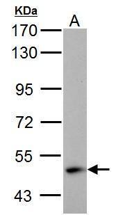
ABAT antibody detects ABAT protein by western blot analysis. A. 30 µg SK-N-SH whole cell lysate/extract 7.5% SDS-PAGE ABAT antibody dilution: 1:1000 The HRP-conjugated anti-rabbit IgG antibody was used to detect the primary antibody.Application
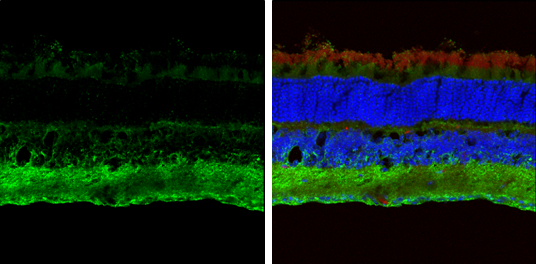
ABAT antibody detects ABAT protein expression by immunohistochemical analysis. Sample: Frozen sectioned adult mouse retina. Green: ABAT protein stained by ABAT antibody diluted at 1:250. Red: beta Tubulin 3/ TUJ1, stained by beta Tubulin 3/ TUJ1 antibody [GT11710] diluted at 1:250. Blue: Fluoroshield with DAPI .Application
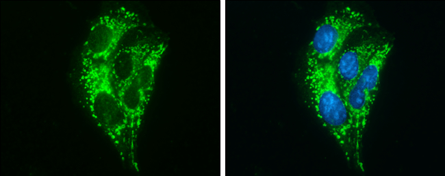
ABAT antibody detects ABAT protein at mitochondria by immunofluorescent analysis. Sample: HepG2 cells were fixed in ice-cold MeOH for 5 min. Green: ABAT protein stained by ABAT antibody diluted at 1:500. Blue: Hoechst 33342 staining.| Product Name | ABAT antibody |
|---|---|
| Antibody Type | Primary Antibodies |
| Product description | Rabbit Polyclonal antibody to ABAT (4-aminobutyrate aminotransferase)4-aminobutyrate aminotransferase (ABAT) is responsible for catabolism of gamma-aminobutyric acid (GABA), an important, mostly inhibitory neurotransmitter in the central nervous system, into succinic semialdehyde. The active enzyme is a homodimer of 50-kD subunits complexed to pyridoxal-5-phosphate. The protein sequence is over 95% similar to the pig protein. GABA is estimated to be present in nearly one-third of human synapses. ABAT in liver and brain is controlled by 2 codominant alleles with a frequency in a Caucasian population of 0.56 and 0.44. The ABAT deficiency phenotype includes psychomotor retardation, hypotonia, hyperreflexia, lethargy, refractory seizures, and EEG abnormalities. Multiple alternatively spliced transcript variants encoding the same protein isoform have been found for this gene. [provided by RefSeq] |
| Immunogen | Recombinant protein encompassing a sequence within the center region of human ABAT. The exact sequence is proprietary. |
| Clonality | Polyclonal |
|---|---|
| Isotype | IgG |
| Host Species | Rabbit |
| Tested Applications | ICC/IFIHC-FIHC-PWB |
| *Optimal dilutions/concentrations should be determined by the researcher.
: |
|
| Species Reactivity | HumanMouseRat |
| Predicted Reactivity | Pig, BovinePig (93%), Bovine (95%) |
| Concentration | 1mg/ml (Please refer to the vial label for the specific concentration) |
| Purification | Affinity purified |
| Gene Synonyms | FLJ17813 antibody FLJ30272 antibody GABA-AT antibody GABAT antibody NPD009 antibody ABAT antibody GABA transaminase antibody 4-aminobutyrate aminotransferase mitochondrial antibody (S)-3-amino-2-methylpropionate transaminase antibody gamma-amino- |
|---|---|
| Molecular Weight(MW) | 56 kDa (note) (3) |
| Cellular Localization | Mitochondrion matrix |
Application
Various tissue extracts (50 µg) were separated by 10% SDS-PAGE, and the membrane was blotted with ABAT antibody diluted at 1:10000. The HRP-conjugated anti-rabbit IgG antibody was used to detect the primary antibody.
Application
ABAT antibody detects ABAT protein at mitochondria on mouse liver by immunohistochemical analysis. Sample: Paraffin-embedded mouse liver. ABAT antibody dilution: 1:500.
Application
ABAT antibody detects ABAT protein at mitochondria on mouse kidney by immunohistochemical analysis. Sample: Paraffin-embedded mouse kidney. ABAT antibody dilution: 1:500.
Application
ABAT antibody detects ABAT protein at cytoplasm in rat brain by immunohistochemical analysis. Sample: Paraffin-embedded rat brain. ABAT antibody diluted at 1:500.
Application
ABAT antibody detects ABAT protein expression by immunohistochemical analysis. Sample: Frozen sectioned E13.5 Rat brain. Green: ABAT protein stained by ABAT antibody diluted at 1:250. Red: beta Tubulin 3/ TUJ1, a mature neuron marker, stained by beta Tubulin 3/ TUJ1 antibody [GT11710] diluted at 1:500. Blue: Fluoroshield with DAPI .
Application
ABAT antibody detects ABAT protein by western blot analysis. A. 30 µg SK-N-SH whole cell lysate/extract 7.5% SDS-PAGE ABAT antibody dilution: 1:1000 The HRP-conjugated anti-rabbit IgG antibody was used to detect the primary antibody.
Application
ABAT antibody detects ABAT protein expression by immunohistochemical analysis. Sample: Frozen sectioned adult mouse retina. Green: ABAT protein stained by ABAT antibody diluted at 1:250. Red: beta Tubulin 3/ TUJ1, stained by beta Tubulin 3/ TUJ1 antibody [GT11710] diluted at 1:250. Blue: Fluoroshield with DAPI .
Application
ABAT antibody detects ABAT protein at mitochondria by immunofluorescent analysis. Sample: HepG2 cells were fixed in ice-cold MeOH for 5 min. Green: ABAT protein stained by ABAT antibody diluted at 1:500. Blue: Hoechst 33342 staining.| Positive Control | SK-N-SH , mouse brain , rat brain |
|---|---|
| Application Notes | *Optimal dilutions/concentrations should be determined by the researcher.
: |
| Form | Liquid |
|---|---|
| Storage Instructions | Keep as concentrated solution. Aliquot and store at -20ºC or below. Avoid multiple freeze-thaw cycles. |
| Storage Buffer | 1XPBS, 1% BSA, 20% Glycerol (pH7). 0.01% Thimerosal was added as a preservative. |


