Application
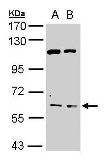
Sample (30 µg of whole cell lysate) A: HepG2 B: Molt-4 7.5% SDS PAGE diluted at 1:500 The HRP-conjugated anti-rabbit IgG antibody was used to detect the primary antibody.Application
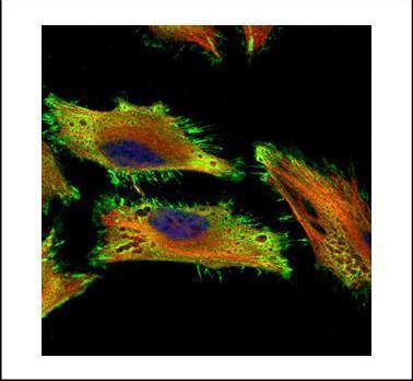
Confocal immunofluorescence analysis (Olympus FV10i) of paraformaldehyde-fixed HeLa, using AChE antibody (Green) at 1:500 dilution. Alpha-tubulin filaments were labeled with (Red) at 1:2000.Application
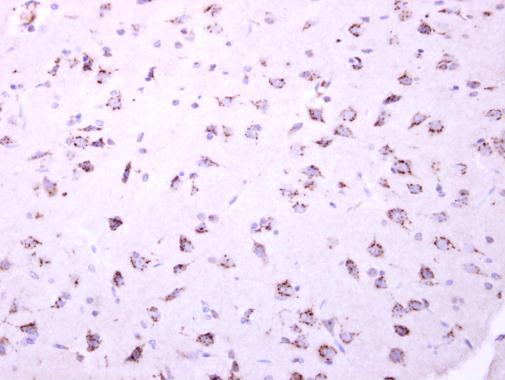
AChE antibody [C3], C-term detects AChE protein at cytoplasm dot-like staining on mouse fore brain by immunohistochemical analysis. Sample: Paraffin-embedded mouse fore brain. AChE antibody [C3], C-term dilution: 1:250.| Product Name | AChE antibody , C-term |
|---|---|
| Antibody Type | Primary Antibodies |
| Product description | Rabbit Polyclonal antibody to AChE (acetylcholinesterase)Acetylcholinesterase hydrolyzes the neurotransmitter, acetylcholine at neuromuscular junctions and brain cholinergic synapses, and thus terminates signal transmission. It is also found on the red blood cell membranes, where it constitutes the Yt blood group antigen. Acetylcholinesterase exists in multiple molecular forms which possess similar catalytic properties, but differ in their oligomeric assembly and mode of cell attachment to the cell surface. It is encoded by the single ACHE gene, and the structural diversity in the gene products arises from alternative mRNA splicing, and post-translational associations of catalytic and structural subunits. The major form of acetylcholinesterase found in brain, muscle and other tissues is the hydrophilic species, which forms disulfide-linked oligomers with collagenous, or lipid-containing structural subunits. The other, alternatively spliced form, expressed primarily in the erythroid tissues, differs at the C-terminal end, and contains a cleavable hydrophobic peptide with a GPI-anchor site. It associates with the membranes through the phosphoinositide (PI) moieties added post-translationally. [provided by RefSeq] |
| Immunogen | Carrier-protein conjugated synthetic peptide encompassing a sequence within the C-terminus region of human AChE. The exact sequence is proprietary. |
| Clonality | Polyclonal |
|---|---|
| Isotype | IgG |
| Host Species | Rabbit |
| Tested Applications | ICC/IFIHC-PWB |
| *Optimal dilutions/concentrations should be determined by the researcher.
: |
|
| Species Reactivity | HumanMouseRat |
| Predicted Reactivity | Cat, Rhesus Monkey, Bovine, Guinea pigCat (100%), Rhesus Monkey (100%), Bovine (100%), Guinea pig (100%) |
| Concentration | 1mg/ml (Please refer to the vial label for the specific concentration) |
| Purification | Affinity purified |
| Gene Synonyms | ARACHE antibody N-ACHE antibody YT antibody ACHE antibody Yt blood group antibody acetylcholinesterase antibody apoptosis-related acetylcholinesterase antibody |
|---|---|
| Molecular Weight(MW) | 68 kDa (note) (3) |
| Cellular Localization | Cell junction , synapse , Secreted , Cell membrane; Peripheral membrane protein , Isoform T: Nucleus , Isoform H: Cell membrane; Lipid-anchor , GPI-anchor; Extracellular side , Cell junction , synapse , Secreted , Cell membrane , Isoform T: Nucleus , Isof |
Application
Sample (30 µg of whole cell lysate) A: HepG2 B: Molt-4 7.5% SDS PAGE diluted at 1:500 The HRP-conjugated anti-rabbit IgG antibody was used to detect the primary antibody.
Application
Confocal immunofluorescence analysis (Olympus FV10i) of paraformaldehyde-fixed HeLa, using AChE antibody (Green) at 1:500 dilution. Alpha-tubulin filaments were labeled with (Red) at 1:2000.
Application
AChE antibody [C3], C-term detects AChE protein at cytoplasm dot-like staining on mouse fore brain by immunohistochemical analysis. Sample: Paraffin-embedded mouse fore brain. AChE antibody [C3], C-term dilution: 1:250.| Positive Control | 293T , A431 , H1299 , HeLaS3 , HepG2 , Molt-4 , Raji |
|---|---|
| Application Notes | *Optimal dilutions/concentrations should be determined by the researcher.
: |
| Form | Liquid |
|---|---|
| Storage Instructions | Keep as concentrated solution. Aliquot and store at -20ºC or below. Avoid multiple freeze-thaw cycles. |
| Storage Buffer | 1XPBS, 1% BSA, 20% Glycerol (pH7). 0.01% Thimerosal was added as a preservative. |


