Application
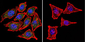
Immunofluorescent analysis of Nicotinic Acetylcholine Receptor using Anti-Nicotinic Acetylcholine Receptor Monoclonal Antibody (88B) shows staining in Hela Cells. Nicotinic Acetylcholine Receptor staining (green), F-Actin staining with Phalloidin (red) and nuclei with DAPI (blue) is shown. Cells were grown on chamber slides and fixed with formaldehyde prior to staining. Cells were probed without (control) or with or an antibody recognizing Nicotinic Acetylcholine Receptor at a dilution of 1:100 over night at 4 C, washed with PBS and incubated with a DyLight-488 conjugated secondary antibody.Application
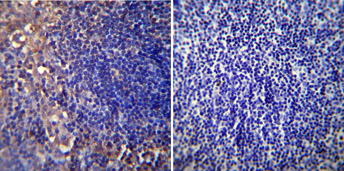
Immunohistochemistry was performed on normal biopsies of de-paraffinized mouse lymph node tissue. To expose target proteins, heat induced antigen retrieval was performed using 10mM sodium citrate (pH6.0) buffer, microwaved for 8-15 minutes. Following antigen retrieval tissues were blocked in 3% BSA-PBS for 30 minutes at room temperature. Tissues were then probed at a dilution of 1:20 with a mouse monoclonal antibody recognizing Nicotinic Acetylcholine Receptor or without primary antibody (negative control) overnight at 4°C in a humidified chamber. Tissues were washed extensively with PBST and endogenous peroxidase activity was quenched with a peroxidase suppressor. Detection was performed using a biotin-conjugated secondary antibody and SA-HRP, followed by colorimetric detection using DAB. Tissues were counterstained with hematoxylin and prepped for mounting.Application
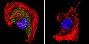
Immunofluorescent analysis of Nicotinic Acetylcholine Receptor using Anti-Nicotinic Acetylcholine Receptor Monoclonal Antibody (88B) shows staining in U251 Cells. Nicotinic Acetylcholine Receptor staining (green), F-Actin staining with Phalloidin (red) and nuclei with DAPI (blue) is shown. Cells were grown on chamber slides and fixed with formaldehyde prior to staining. Cells were probed without (control) or with or an antibody recognizing Nicotinic Acetylcholine Receptor at a dilution of 1:20 over night at 4 C, washed with PBS and incubated with a DyLight-488 conjugated secondary antibody.Application
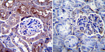
Immunohistochemistry was performed on normal biopsies of de-paraffinized Mouse kidney tissue. To expose target proteins, heat induced antigen retrieval was performed using 10mM sodium citrate (pH 6.0) buffer, microwaved for 8-15 minutes. Following antigen retrieval tissues were blocked in 3% BSA-PBS for 30 minutes at room temperature. Tissues were then probed at a dilution of 1:20 with a mouse monoclonal antibody recognizing Nicotinic Acetylcholine Receptor or without primary antibody (negative control) overnight at 4°C in a humidified chamber. Tissues were washed extensively with PBST and endogenous peroxidase activity was quenched with a peroxidase suppressor. Detection was performed using a biotin-conjugated secondary antibody and SA-HRP, followed by colorimetric detection using DAB. Tissues were counterstained with hematoxylin and prepped for mounting.Application
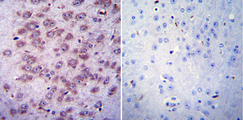
Immunohistochemistry was performed on normal biopsies of de-paraffinized Mouse brain tissue. To expose target proteins, heat induced antigen retrieval was performed using 10mM sodium citrate (pH 6.0) buffer, microwaved for 8-15 minutes. Following antigen retrieval tissues were blocked in 3% BSA-PBS for 30 minutes at room temperature. Tissues were then probed at a dilution of 1:20 with a mouse monoclonal antibody recognizing Nicotinic Acetylcholine Receptor or without primary antibody (negative control) overnight at 4°C in a humidified chamber. Tissues were washed extensively with PBST and endogenous peroxidase activity was quenched with a peroxidase suppressor. Detection was performed using a biotin-conjugated secondary antibody and SA-HRP, followed by colorimetric detection using DAB. Tissues were counterstained with hematoxylin and prepped for mounting.Application
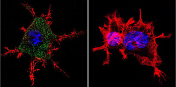
Immunofluorescent analysis of Nicotinic Acetylcholine Receptor using Anti-Nicotinic Acetylcholine Receptor Monoclonal Antibody (88B) shows staining in Neuro-2a Cells. Nicotinic Acetylcholine Receptor staining (green), F-Actin staining with Phalloidin (red) and nuclei with DAPI (blue) is shown. Cells were grown on chamber slides and fixed with formaldehyde prior to staining. Cells were probed without (control) or with or an antibody recognizing Nicotinic Acetylcholine Receptor at a dilution of 1:100 over night at 4 C, washed with PBS and incubated with a DyLight-488 conjugated secondary antibody.| Product Name | AChR antibody |
|---|---|
| Antibody Type | Primary Antibodies |
| Product description | Mouse Monoclonal antibody [88B] to AChRAcetylcholine receptors (AChRs) mediate synaptic transmission at the neuromuscular junction. The channel-linked AChR that mediates rapid, excitatory actions of acetylcholine is called nicotinic AChR (nAChR) because it can be activated by nicotine. The non-channel linked AChR that medicates the slow actions of acetylcholine, which can be either inhibitory or excitatory, is called muscarinic AChR (mAChR) because it can be activated by muscarine. The nAChRs fall into two subclasses, the muscle-type and the neuronal-type. The muscle-type receptor is a pentameric glycoprotein of five membrane spanning subunits, (2 alpha 1’s, beta 1, gamma or epsilon and delta) which form a ligand gated ion channel. The neuronal nAChR is also a pentamer, but unlike the muscle-type, is made up of only two general types of subunits, alpha and beta of which multiple subtypes have been described (alpha 2 to alpha 8 and beta 2 to beta 4). |
| Immunogen | Purified Torpedo californica acetylcholine receptor. |
| Clonality | Monoclonal |
|---|---|
| Isotype | IgG1 |
| Clone Number | 88B |
| Host Species | Mouse |
| Tested Applications | ELISAICC/IFIHCIHC-FIPWB |
| GTX22804 has been successfully used in western blot, immunohistochemistry and immunoprecipitation procedures. By western blot, this antibody detects a 60 kDa protein representing the gamma subunit and a 65 kDa protein representing the delta subunit from torpedo skeletal muscle homogenates. Under non-reducing conditions the delta subunit migrates mostly as a dimer of ~130 kDa. Immunohistochemical staining of nAChR in rat skeletal muscle with GTX22804 results in strong staining of the motor endplate. In Immunohistochemical staining, cryostat or permeabilization of the sections is recommended due to the reactivity of this antibody with the cytoplasmic side of the receptor.: |
|
| Species Reactivity | HumanMouseChickenRatAmphibiansFishRabbitVertebratesTorpedo |
| Concentration | 1mg/ml |
| Purification | Affinity purified |
| Gene Synonyms | CHRND CHNRD |
|---|---|
| Target | AChR |
| Tissue Specificity | detects nicotinic acetylcholine receptor (nAChR) gamma and delta subunits in torpedo and the delta subunit in mouse, human, rat, chicken and frog. This antibody does not detect the alpha 1 and beta 1 subunits. |
Application
Immunofluorescent analysis of Nicotinic Acetylcholine Receptor using Anti-Nicotinic Acetylcholine Receptor Monoclonal Antibody (88B) shows staining in Hela Cells. Nicotinic Acetylcholine Receptor staining (green), F-Actin staining with Phalloidin (red) and nuclei with DAPI (blue) is shown. Cells were grown on chamber slides and fixed with formaldehyde prior to staining. Cells were probed without (control) or with or an antibody recognizing Nicotinic Acetylcholine Receptor at a dilution of 1:100 over night at 4 C, washed with PBS and incubated with a DyLight-488 conjugated secondary antibody.
Application
Immunohistochemistry was performed on normal biopsies of de-paraffinized mouse lymph node tissue. To expose target proteins, heat induced antigen retrieval was performed using 10mM sodium citrate (pH6.0) buffer, microwaved for 8-15 minutes. Following antigen retrieval tissues were blocked in 3% BSA-PBS for 30 minutes at room temperature. Tissues were then probed at a dilution of 1:20 with a mouse monoclonal antibody recognizing Nicotinic Acetylcholine Receptor or without primary antibody (negative control) overnight at 4°C in a humidified chamber. Tissues were washed extensively with PBST and endogenous peroxidase activity was quenched with a peroxidase suppressor. Detection was performed using a biotin-conjugated secondary antibody and SA-HRP, followed by colorimetric detection using DAB. Tissues were counterstained with hematoxylin and prepped for mounting.
Application
Immunofluorescent analysis of Nicotinic Acetylcholine Receptor using Anti-Nicotinic Acetylcholine Receptor Monoclonal Antibody (88B) shows staining in U251 Cells. Nicotinic Acetylcholine Receptor staining (green), F-Actin staining with Phalloidin (red) and nuclei with DAPI (blue) is shown. Cells were grown on chamber slides and fixed with formaldehyde prior to staining. Cells were probed without (control) or with or an antibody recognizing Nicotinic Acetylcholine Receptor at a dilution of 1:20 over night at 4 C, washed with PBS and incubated with a DyLight-488 conjugated secondary antibody.
Application
Immunohistochemistry was performed on normal biopsies of de-paraffinized Mouse kidney tissue. To expose target proteins, heat induced antigen retrieval was performed using 10mM sodium citrate (pH 6.0) buffer, microwaved for 8-15 minutes. Following antigen retrieval tissues were blocked in 3% BSA-PBS for 30 minutes at room temperature. Tissues were then probed at a dilution of 1:20 with a mouse monoclonal antibody recognizing Nicotinic Acetylcholine Receptor or without primary antibody (negative control) overnight at 4°C in a humidified chamber. Tissues were washed extensively with PBST and endogenous peroxidase activity was quenched with a peroxidase suppressor. Detection was performed using a biotin-conjugated secondary antibody and SA-HRP, followed by colorimetric detection using DAB. Tissues were counterstained with hematoxylin and prepped for mounting.
Application
Immunohistochemistry was performed on normal biopsies of de-paraffinized Mouse brain tissue. To expose target proteins, heat induced antigen retrieval was performed using 10mM sodium citrate (pH 6.0) buffer, microwaved for 8-15 minutes. Following antigen retrieval tissues were blocked in 3% BSA-PBS for 30 minutes at room temperature. Tissues were then probed at a dilution of 1:20 with a mouse monoclonal antibody recognizing Nicotinic Acetylcholine Receptor or without primary antibody (negative control) overnight at 4°C in a humidified chamber. Tissues were washed extensively with PBST and endogenous peroxidase activity was quenched with a peroxidase suppressor. Detection was performed using a biotin-conjugated secondary antibody and SA-HRP, followed by colorimetric detection using DAB. Tissues were counterstained with hematoxylin and prepped for mounting.
Application
Immunofluorescent analysis of Nicotinic Acetylcholine Receptor using Anti-Nicotinic Acetylcholine Receptor Monoclonal Antibody (88B) shows staining in Neuro-2a Cells. Nicotinic Acetylcholine Receptor staining (green), F-Actin staining with Phalloidin (red) and nuclei with DAPI (blue) is shown. Cells were grown on chamber slides and fixed with formaldehyde prior to staining. Cells were probed without (control) or with or an antibody recognizing Nicotinic Acetylcholine Receptor at a dilution of 1:100 over night at 4 C, washed with PBS and incubated with a DyLight-488 conjugated secondary antibody.| Application Notes | GTX22804 has been successfully used in western blot, immunohistochemistry and immunoprecipitation procedures. By western blot, this antibody detects a 60 kDa protein representing the gamma subunit and a 65 kDa protein representing the delta subunit from torpedo skeletal muscle homogenates. Under non-reducing conditions the delta subunit migrates mostly as a dimer of ~130 kDa. Immunohistochemical staining of nAChR in rat skeletal muscle with GTX22804 results in strong staining of the motor endplate. In Immunohistochemical staining, cryostat or permeabilization of the sections is recommended due to the reactivity of this antibody with the cytoplasmic side of the receptor.: |
|---|
| Form | Liquid |
|---|---|
| Storage Instructions | Keep as concentrated solution. Aliquot and store at -80ºC or below. Avoid multiple freeze-thaw cycles. |
| Storage Buffer | PBS containing 0.05% sodium azide. |


