Application
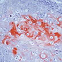
Formalin-fixed, paraffin-embedded human squamous cell carcinoma of lung stained with using peroxidase-conjugate and AEC chromogen. Note cytoplasmic staining of tumor cells.| Product Name | Acidic Cytokeratin antibody antibody |
|---|---|
| Antibody Type | Primary Antibodies |
| Product description | Mouse monoclonal antibody [AE1] to Acidic CytokeratinThis gene encodes a member of the type I (acidic) cytokeratin family, which belongs to the superfamily of intermediate filament (IF) proteins. Keratins are heteropolymeric structural proteins which form the intermediate filament. These filaments, along with actin microfilaments and microtubules, compose the cytoskeleton of epithelial cells. Mutations in this gene are associated with epidermolytic hyperkeratosis. This gene is located within a cluster of keratin family members on chromosome 17q21. [provided by RefSeq, Jul 2008] |
| Immunogen | Human epidermal keratin |
| Clonality | Monoclonal |
|---|---|
| Isotype | IgG1 |
| Clone Number | AE1 |
| Host Species | Mouse |
| Tested Applications | ICC/IFIHC-P |
| NOT Suitable for use in Western blotting. For staining of formalin-fixed tissues, digest sections with trypsin at 1mg/ml PBS, 10 min at 37¡C or Protease XXV at 1mg/ml PBS for 5minutes at 37¡C. This antibody is an excellent marker for distinguishing carcinomas from non-epithelial tumours. This antibody used alongside GTX29287 has also been used extensively for distinguishing carcinoma from non-carcinoma. Optimal dilutions/concentrations should be determined by the researcher.: |
|
| Species Reactivity | HumanMouseMonkeyRatRabbitBovine |
| Concentration | 0.2mg/ml |
| Purification | Affinity purified |
| Gene Synonyms | KRT10 BIE K10 KPP EHK CK10 BCIE |
|---|---|
| Target | Acidic Cytokeratin |
| Tissue Specificity | This antibody reacts with MCF-7, RT 112 and A 431 cultured cell lines. |
| Cellular Localization | Cytoplasmic. |
Application
Formalin-fixed, paraffin-embedded human squamous cell carcinoma of lung stained with using peroxidase-conjugate and AEC chromogen. Note cytoplasmic staining of tumor cells.| Positive Control | Skin, Squamous cellcarcinoma |
|---|---|
| Application Notes | NOT Suitable for use in Western blotting. For staining of formalin-fixed tissues, digest sections with trypsin at 1mg/ml PBS, 10 min at 37¡C or Protease XXV at 1mg/ml PBS for 5minutes at 37¡C. This antibody is an excellent marker for distinguishing carcinomas from non-epithelial tumours. This antibody used alongside GTX29287 has also been used extensively for distinguishing carcinoma from non-carcinoma. Optimal dilutions/concentrations should be determined by the researcher.: |
| Form | Liquid |
|---|---|
| Storage Instructions | Keep as concentrated solution, aliquot and store at 4°C. Do not freeze. |
| Storage Buffer | 20 mM Tris-Borate, 150 mM sodium chloride, dialyzed media RPMI 1640/D-MEM containing fetal bovine serum, BMC-6 Carrier Polysaccharides, Carrier Protein, and 0.05% sodium azide, pH 7.5. |


