Application
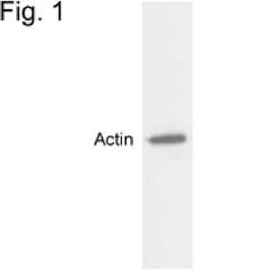
Western blot detection of actin in MDCK cell extract.Application
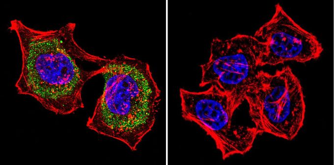
ICC/IF analysis of HeLa cells using Actin antibody at a dilution of 1:20 (green). F-Actin staining with Phalloidin (red) and nuclei with DAPI (blue) is shown. Images were taken at 60X magnification.Application
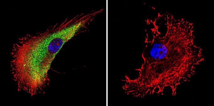
Immunofluorescent analysis of Actin in A375 cells. Actin staining (green), F-Actin staining with Phalloidin (red) and nuclei with DAPI (blue) is shown. Cells were grown on slides and fixed with formaldehyde prior to staining. Cells were probed without (control) or with Actin antibody [mAbGEa] at a dilution of 1:200 over night at 4 ?C, washed with PBS and incubated with a proper secondary antibody. Images were taken at 60X magnification.Application
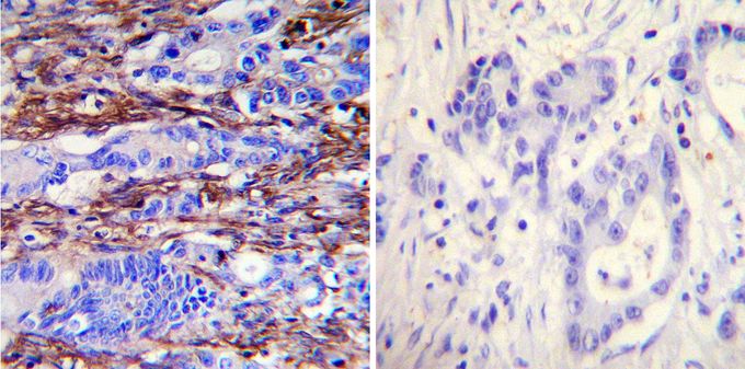
Immunohistochemistry was performed on cancer biopsies of deparaffinized human colon carcinoma tissues. To expose target proteins, heat induced antigen retrieval was performed using 10mM sodium citrate (pH6.0) buffer, microwaved for 8-15 minutes. Following antigen retrieval tissues were blocked in 3% BSA-PBS for 30 minutes at room temperature. Tissues were then probed at a dilution of 1:100 with or without Actin antibody [mAbGEa] overnight at 4°C in a humidified chamber. Tissues were washed extensively with PBST and endogenous peroxidase activity was quenched with a peroxidase suppressor. Detection was performed using a biotin-conjugated secondary antibody and SA-HRP, followed by colorimetric detection using DAB. Tissues were counterstained with hematoxylin and prepped for mounting.| Product Name | Actin antibody |
|---|---|
| Antibody Type | Primary Antibodies |
| Product description | Mouse monoclonal antibody [mAbGEa] to ActinActins are highly conserved proteins that are involved in various types of cell motility. Polymerization of globular actin (G-actin) leads to a structural filament (F-actin) in the form of a two-stranded helix. Each actin can bind to four others. The protein encoded by this gene belongs to the actin family which is comprised of three main groups of actin isoforms, alpha, beta, and gamma. The alpha actins are found in muscle tissues and are a major constituent of the contractile apparatus. Defects in this gene have been associated with idiopathic dilated cardiomyopathy (IDC) and familial hypertrophic cardiomyopathy (FHC). [provided by RefSeq, Jul 2008] |
| Immunogen | Purified Arabidopsis actin protein. |
| Conjugation | Agarose |
| Modification | Acetylated |
| Clonality | Monoclonal |
|---|---|
| Isotype | IgG1 |
| Clone Number | mAbGEa |
| Host Species | Mouse |
| Tested Applications | ELISAICC/IFIHCIHC-PWB |
| WB:1:500~1:2000 ICC/IF:1:50~1:100 IHC:1:50~1:200 |
|
| Species Reactivity | BacteriaBovineDogDrosophilaHamsterHumanMousePigPlantRatSheepXenopusYeastZebra Fish |
| Concentration | 1mg/ml |
| Purification | Affinity purified |
| Gene Synonyms | CMH11 LVNC4 ACTC1 ASD5 ACTC actin alpha cardiac muscle 1 CMD1R |
|---|---|
| Target | Actin |
| Tissue Specificity | This antibody has shown cross-reactivity with actin 1, 2, 3, 4, 7, 8, 11 and 12. |
Application
Western blot detection of actin in MDCK cell extract.
Application
ICC/IF analysis of HeLa cells using Actin antibody at a dilution of 1:20 (green). F-Actin staining with Phalloidin (red) and nuclei with DAPI (blue) is shown. Images were taken at 60X magnification.
Application
Immunofluorescent analysis of Actin in A375 cells. Actin staining (green), F-Actin staining with Phalloidin (red) and nuclei with DAPI (blue) is shown. Cells were grown on slides and fixed with formaldehyde prior to staining. Cells were probed without (control) or with Actin antibody [mAbGEa] at a dilution of 1:200 over night at 4 ?C, washed with PBS and incubated with a proper secondary antibody. Images were taken at 60X magnification.
Application
Immunohistochemistry was performed on cancer biopsies of deparaffinized human colon carcinoma tissues. To expose target proteins, heat induced antigen retrieval was performed using 10mM sodium citrate (pH6.0) buffer, microwaved for 8-15 minutes. Following antigen retrieval tissues were blocked in 3% BSA-PBS for 30 minutes at room temperature. Tissues were then probed at a dilution of 1:100 with or without Actin antibody [mAbGEa] overnight at 4°C in a humidified chamber. Tissues were washed extensively with PBST and endogenous peroxidase activity was quenched with a peroxidase suppressor. Detection was performed using a biotin-conjugated secondary antibody and SA-HRP, followed by colorimetric detection using DAB. Tissues were counterstained with hematoxylin and prepped for mounting.| Application Notes | WB:1:500~1:2000 ICC/IF:1:50~1:100 IHC:1:50~1:200 |
|---|
| Form | Liquid |
|---|---|
| Storage Instructions | -20° C, Avoid Freeze/Thaw Cycles |
| Storage Buffer | Ascites containing 0.05 % sodium azide |


