Application
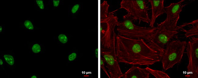
ADAR1 antibody [N3C1], Internal detects ADAR1 protein at nucleolus and nucleus by immunofluorescent analysis. Sample: HeLa cells were fixed in 4% paraformaldehyde at RT for 15 min. Green: ADAR1 protein stained by ADAR1 antibody [N3C1], Internal diluted at 1:200. Red: phalloidin, a cytoskeleton marker, diluted at 1:200. Blue: Hoechst 33342 staining. Scale bar = 10 μm.Application
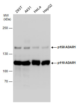
Various whole cell extracts (30 µg) were separated by 5% SDS-PAGE, and the membrane was blotted with ADAR1 antibody [N3C1], Internal diluted at 1:1000. The HRP-conjugated anti-rabbit IgG antibody was used to detect the primary antibody.Application
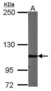
Sample (30 µg of whole cell lysate) A:NIH-3T3 5% SDS PAGE diluted at 1:1000 The HRP-conjugated anti-rabbit IgG antibody was used to detect the primary antibody.Application
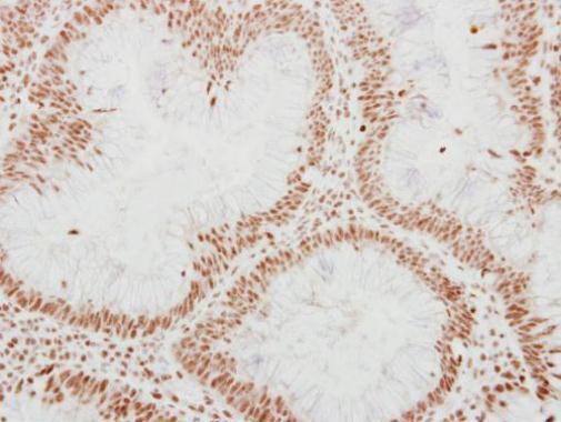
Immunohistochemical analysis of paraffin-embedded human colon carcinoma, using ADAR1 antibody at 1:250 dilution.Application
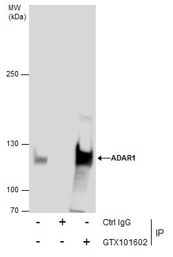
Immunoprecipitation of ADAR1 protein from 293T whole cell extracts using 5 μg of ADAR1 antibody [N3C1], Internal . Western blot analysis was performed using ADAR1 antibody [N3C1], Internal . EasyBlot anti-Rabbit IgG was used as a secondary reagent.Application
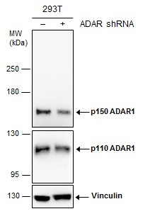
Non-transfected (–) and transfected (+) 293T whole cell extracts (30 µg) were separated by 5% SDS-PAGE, and the membrane was blotted with ADAR1 antibody [N3C1], Internal diluted at 1:3000. The HRP-conjugated anti-rabbit IgG antibody was used to detect the primary antibody.| Product Name | ADAR1 antibody , Internal |
|---|---|
| Antibody Type | Primary Antibodies |
| Product description | Rabbit Polyclonal antibody to ADAR1 (adenosine deaminase, RNA-specific)This gene encodes the enzyme responsible for RNA editing by site-specific deamination of adenosines. This enzyme destabilizes double stranded RNA through conversion of adenosine to inosine. Mutations in this gene have been associated with dyschromatosis symmetrica hereditaria. Alternate transcriptional splice variants, encoding different isoforms, have been characterized. [provided by RefSeq] |
| Immunogen | Recombinant protein encompassing a sequence within the center region of human ADAR1. The exact sequence is proprietary. |
| Clonality | Polyclonal |
|---|---|
| Isotype | IgG |
| Host Species | Rabbit |
| Tested Applications | ICC/IFIHC-PIPWB |
| *Optimal dilutions/concentrations should be determined by the researcher.
: |
|
| Species Reactivity | HumanMouse |
| Predicted Reactivity | RatRat (80%) |
| Concentration | 0.5mg/ml |
| Purification | Affinity purified |
| Gene Synonyms | ADAR1 antibody DRADA antibody DSH antibody DSRAD antibody G1P1 antibody IFI-4 antibody IFI4 antibody K88DSRBP antibody P136 antibody ADAR antibody 136 kDa double-stranded RNA-binding protein antibody dsRNA adenosine deaminase antibody interfero |
|---|---|
| Molecular Weight(MW) | 136 kDa (note) (5) |
| Cellular Localization | Cytoplasm , Nucleus , nucleolus |
Application
ADAR1 antibody [N3C1], Internal detects ADAR1 protein at nucleolus and nucleus by immunofluorescent analysis. Sample: HeLa cells were fixed in 4% paraformaldehyde at RT for 15 min. Green: ADAR1 protein stained by ADAR1 antibody [N3C1], Internal diluted at 1:200. Red: phalloidin, a cytoskeleton marker, diluted at 1:200. Blue: Hoechst 33342 staining. Scale bar = 10 μm.
Application
Various whole cell extracts (30 µg) were separated by 5% SDS-PAGE, and the membrane was blotted with ADAR1 antibody [N3C1], Internal diluted at 1:1000. The HRP-conjugated anti-rabbit IgG antibody was used to detect the primary antibody.
Application
Sample (30 µg of whole cell lysate) A:NIH-3T3 5% SDS PAGE diluted at 1:1000 The HRP-conjugated anti-rabbit IgG antibody was used to detect the primary antibody.
Application
Immunohistochemical analysis of paraffin-embedded human colon carcinoma, using ADAR1 antibody at 1:250 dilution.
Application
Immunoprecipitation of ADAR1 protein from 293T whole cell extracts using 5 μg of ADAR1 antibody [N3C1], Internal . Western blot analysis was performed using ADAR1 antibody [N3C1], Internal . EasyBlot anti-Rabbit IgG was used as a secondary reagent.
Application
Non-transfected (–) and transfected (+) 293T whole cell extracts (30 µg) were separated by 5% SDS-PAGE, and the membrane was blotted with ADAR1 antibody [N3C1], Internal diluted at 1:3000. The HRP-conjugated anti-rabbit IgG antibody was used to detect the primary antibody.| Positive Control | 293T , A431 , HeLa , HepG2 , NIH-3T3 |
|---|---|
| Application Notes | *Optimal dilutions/concentrations should be determined by the researcher.
: |
| Form | Liquid |
|---|---|
| Storage Instructions | Keep as concentrated solution. Aliquot and store at -20ºC or below. Avoid multiple freeze-thaw cycles. |
| Storage Buffer | 1XPBS, 1% BSA, 20% Glycerol (pH7). 0.01% Thimerosal was added as a preservative. |


