Application
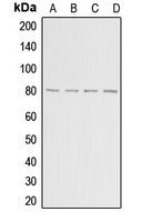
Western blot analysis of ADAR2 expression in HepG2 (A); MCF7 (B); mouse brain (C); rat brain (D) whole cell lysates.Application
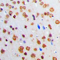
Immunohistochemical analysis of ADAR2 staining in human brain formalin fixed paraffin embedded tissue section. The section was pre-treated using heat mediated antigen retrieval with sodium citrate buffer (pH 6.0). The section was then incubated with the antibody at room temperature and detected using an HRP conjugated compact polymer system. DAB was used as the chromogen. The section was then counterstained with haematoxylin and mounted with DPX.Application
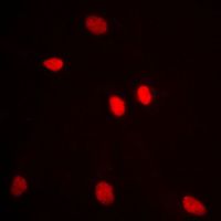
Immunofluorescent analysis of ADAR2 staining in MCF7 cells. Formalin-fixed cells were permeabilized with 0.1% Triton X-100 in TBS for 5-10 minutes and blocked with 3% BSA-PBS for 30 minutes at room temperature. Cells were probed with the primary antibody in 3% BSA-PBS and incubated overnight at 4 C in a humidified chamber. Cells were washed with PBST and incubated with a DyLight 594-conjugated secondary antibody (red) in PBS at room temperature in the dark. DAPI was used to stain the cell nuclei (blue).| Product Name | ADAR2 antibody |
|---|---|
| Antibody Type | Primary Antibodies |
| Product description | Rabbit Polyclonal antibody to ADAR2This gene encodes the enzyme responsible for pre-mRNA editing of the glutamate receptor subunit B by site-specific deamination of adenosines. Studies in rat found that this enzyme acted on its own pre-mRNA molecules to convert an AA dinucleotide to an AI dinucleotide which resulted in a new splice site. Alternative splicing of this gene results in several transcript variants, some of which have been characterized by the presence or absence of an ALU cassette insert and a short or long C-terminal region. [provided by RefSeq, Jul 2008] |
| Immunogen | The immunogen corresponding to a region within amino acids 496 and 535 of human ADAR2. |
| Clonality | Polyclonal |
|---|---|
| Isotype | IgG |
| Host Species | Rabbit |
| Tested Applications | ICC/IFIHC-PWB |
| WB: 1:500 - 1:1000. IHC-P: 1:100 - 1:200. ICC/IF: 1:100 - 1:500: |
|
| Species Reactivity | HumanMouseRat |
| Concentration | 1mg/ml |
| Purification | Affinity purified |
| Gene Synonyms | ADARB1 DRABA2 RED1 DRADA2 |
|---|---|
| Target | ADAR2 |
Application
Western blot analysis of ADAR2 expression in HepG2 (A); MCF7 (B); mouse brain (C); rat brain (D) whole cell lysates.
Application
Immunohistochemical analysis of ADAR2 staining in human brain formalin fixed paraffin embedded tissue section. The section was pre-treated using heat mediated antigen retrieval with sodium citrate buffer (pH 6.0). The section was then incubated with the antibody at room temperature and detected using an HRP conjugated compact polymer system. DAB was used as the chromogen. The section was then counterstained with haematoxylin and mounted with DPX.
Application
Immunofluorescent analysis of ADAR2 staining in MCF7 cells. Formalin-fixed cells were permeabilized with 0.1% Triton X-100 in TBS for 5-10 minutes and blocked with 3% BSA-PBS for 30 minutes at room temperature. Cells were probed with the primary antibody in 3% BSA-PBS and incubated overnight at 4 C in a humidified chamber. Cells were washed with PBST and incubated with a DyLight 594-conjugated secondary antibody (red) in PBS at room temperature in the dark. DAPI was used to stain the cell nuclei (blue).| Application Notes | WB: 1:500 - 1:1000. IHC-P: 1:100 - 1:200. ICC/IF: 1:100 - 1:500: |
|---|
| Form | Liquid |
|---|---|
| Storage Instructions | Shipped at 4 C. Upon delivery aliquot and store at -20 C for one year. Avoid freeze/thaw cycles. |
| Storage Buffer | Liquid in 0.42% Potassium phosphate, 0.87% Sodium chloride, pH 7.3, 30% glycerol and 0.01% sodium azide. |


