Application
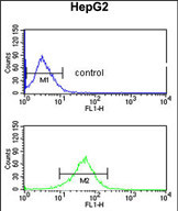
AGPAT3 Antibody (Center) flow cytometric analysis of HepG2 cells (bottom histogram) compared to a negative control cell (top histogram).FITC-conjugated goat-anti-rabbit secondary antibodies were used for the analysis.Application
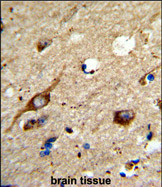
Formalin-fixed and paraffin-embedded human brain tissue reacted with AGPAT3 Antibody (Center), which was peroxidase-conjugated to the secondary antibody, followed by DAB staining. This data demonstrates the use of this antibody for immunohistochemistry; clinical relevance has not been evaluated.| Product Name | AGPAT3 antibody, Internal |
|---|---|
| Antibody Type | Primary Antibodies |
| Product description | Rabbit polyclonal to AGPAT3 (Internal)The protein encoded by this gene is an acyltransferase that converts lysophosphatidic acid into phosphatidic acid, which is the second step in the de novo phospholipid biosynthetic pathway. The encoded protein may be an integral membrane protein. Two transcript variants encoding the same protein have been found for this gene. [provided by RefSeq] |
| Immunogen | KLH conjugated synthetic peptide between 248~277 amino acids from the Center region of human AGPAT3. |
| Clonality | Polyclonal |
|---|---|
| Isotype | IgG |
| Host Species | Rabbit |
| Tested Applications | FACSIHC-P |
| ELISA: Use at 1:1,000 | |
| Species Reactivity | Human |
| Concentration | 0.2mg/ml |
| Purification | Affinity purified |
| Gene Synonyms | 1acylsnglycerol3phosphate acyltransferase gamma, 1AGPAT 3, 1AGP acyltransferase 3, LPAATgamma, 1AGPAT3, AGPAT3, 1-acylglycerol-3-phosphate O-acyltransferase 3, 1-AGP acyltransferase 3, 1acylglycerol3phosphate Oacyltransferase 3, LPAAT-gamma, 1AGP acyltrans |
|---|---|
| Target | AGPAT3 |
| Molecular Weight(MW) | 43.381 kDa (note) |
Application
AGPAT3 Antibody (Center) flow cytometric analysis of HepG2 cells (bottom histogram) compared to a negative control cell (top histogram).FITC-conjugated goat-anti-rabbit secondary antibodies were used for the analysis.
Application
Formalin-fixed and paraffin-embedded human brain tissue reacted with AGPAT3 Antibody (Center), which was peroxidase-conjugated to the secondary antibody, followed by DAB staining. This data demonstrates the use of this antibody for immunohistochemistry; clinical relevance has not been evaluated.| Application Notes | ELISA: Use at 1:1,000 |
|---|
| Form | Liquid |
|---|---|
| Storage Instructions | Store at at 2-8°C for up to 6 months. For long term storage, aliquot and store at -20 °C. Avoid repeated freeze/thaw cycles. |
| Storage Buffer | PBS with 0.09% (W/V) sodium azide |


