Application
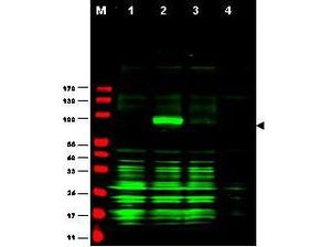
Western blot using Affinity Purified anti-Ajuba antibody shows detection of Ajuba-RFP fusion protein in cell lysates (arrow-head). Lanes correspond to 1) vector only trans-fection, 2) human Ajuba-RFP, 3) mouse Ajuba-RFP, and 4) mock transfection. Approximately 50 µg of each lysate was loaded per lane for SDS-PAGE followed by transfer onto nitrocellulose and reaction with a 1:1,700 dilution of anti-Ajuba antibody.Application
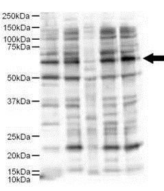
Western blot using Affinity Purified anti-Ajuba antibody shows detection of a 57-kDa band consistent with the expected MW for Ajuba (arrowhead). Lanes correspond to 1) HeLa nuclear extract, and 2) HeLa, 3) A431, 4) Jurkat and 5) 293 whole cell lysates. Immunoprecipitation of Ajuba followed by western blotting may result in cleaner background staining. Approximately 5 µg of each preparation was run on a SDS-PAGE and transferred onto nitrocellulose followed by reaction with a 1:500 dilution of anti-Ajuba antibody. Detection occurred using a 1:5,000 dilution of HRP-labeled Donkey anti-Rabbit IgG for 1 hour at room temperature. A chemiluminescence system was used for signal detection (Roche) using a 60-sec exposure time.| Product Name | Ajuba antibody |
|---|---|
| Antibody Type | Primary Antibodies |
| Product description | Rabbit Polyclonal antibody to AjubaHuman Ajuba (also called JUB protein and ajuba homolog isoform 1) is a LIM domain protein suggested to bind and regulate the activity of Aurora A. Aurora A, which is involved in cell cycle regulation, is upregulated during mitosis, localizing to the centrosomes and microtubule regions proximal to the centrosomes. |
| Immunogen | This affinity purified antibody was prepared from whole rabbit serum produced by repeated immunizations with a synthetic peptide corresponding aa 224-239 of Human Ajuba. |
| Clonality | Polyclonal |
|---|---|
| Isotype | IgG |
| Host Species | Rabbit |
| Tested Applications | ELISAIPWB |
| *Optimal dilutions/concentrations should be determined by the researcher. | |
| Species Reactivity | Human |
| Concentration | 1mg/ml |
| Purification | Affinity purified |
| Gene Synonyms | AJUBA , JUB |
|---|---|
| Target | Ajuba |
| Cellular Localization | Shuttles between the cytoplasm and the nucleus. Localizes on centrosomes during G2-M phase. Preferentially co-localizes with cadherin-adhesive complexes at sites of cell-cell contacts. |
Application
Western blot using Affinity Purified anti-Ajuba antibody shows detection of Ajuba-RFP fusion protein in cell lysates (arrow-head). Lanes correspond to 1) vector only trans-fection, 2) human Ajuba-RFP, 3) mouse Ajuba-RFP, and 4) mock transfection. Approximately 50 µg of each lysate was loaded per lane for SDS-PAGE followed by transfer onto nitrocellulose and reaction with a 1:1,700 dilution of anti-Ajuba antibody.
Application
Western blot using Affinity Purified anti-Ajuba antibody shows detection of a 57-kDa band consistent with the expected MW for Ajuba (arrowhead). Lanes correspond to 1) HeLa nuclear extract, and 2) HeLa, 3) A431, 4) Jurkat and 5) 293 whole cell lysates. Immunoprecipitation of Ajuba followed by western blotting may result in cleaner background staining. Approximately 5 µg of each preparation was run on a SDS-PAGE and transferred onto nitrocellulose followed by reaction with a 1:500 dilution of anti-Ajuba antibody. Detection occurred using a 1:5,000 dilution of HRP-labeled Donkey anti-Rabbit IgG for 1 hour at room temperature. A chemiluminescence system was used for signal detection (Roche) using a 60-sec exposure time.| Application Notes | *Optimal dilutions/concentrations should be determined by the researcher. |
|---|
| Form | Liquid |
|---|---|
| Storage Instructions | Store vial at -20° C prior to opening. Aliquot contents and freeze at -20° C or below for extended storage. Avoid cycles of freezing and thawing. |
| Storage Buffer | 0.02 M Potassium Phosphate, 0.15 M Sodium Chloride, pH 7.2 and 0.01% (w/v) Sodium Azide |


