Application
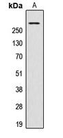
Western blot analysis of AKAP13 expression in HeLa (A) whole cell lysates.Application
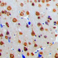
Immunohistochemical analysis of AKAP13 staining in human brain formalin fixed paraffin embedded tissue section. The section was pre-treated using heat mediated antigen retrieval with sodium citrate buffer (pH 6.0). The section was then incubated with the antibody at room temperature and detected using an HRP conjugated compact polymer system. DAB was used as the chromogen. The section was then counterstained with haematoxylin and mounted with DPX.Application
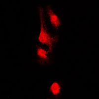
Immunofluorescent analysis of AKAP13 staining in HeLa cells. Formalin-fixed cells were permeabilized with 0.1% Triton X-100 in TBS for 5-10 minutes and blocked with 3% BSA-PBS for 30 minutes at room temperature. Cells were probed with the primary antibody in 3% BSA-PBS and incubated overnight at 4 C in a humidified chamber. Cells were washed with PBST and incubated with a DyLight 594-conjugated secondary antibody (red) in PBS at room temperature in the dark. DAPI was used to stain the cell nuclei (blue).| Product Name | AKAP13 antibody |
|---|---|
| Antibody Type | Primary Antibodies |
| Product description | Rabbit Polyclonal antibody to AKAP13The A-kinase anchor proteins (AKAPs) are a group of structurally diverse proteins which have the common function of binding to the regulatory subunit of protein kinase A (PKA) and confining the holoenzyme to discrete locations within the cell. This gene encodes a member of the AKAP family. Alternative splicing of this gene results in multiple transcript variants encoding different isoforms containing c-terminal dbl oncogene homology (DH) and pleckstrin homology (PH) domains. The DH domain is associated with guanine nucleotide exchange activation for the Rho/Rac family of small GTP binding proteins, resulting in the conversion of the inactive GTPase to the active form capable of transducing signals. The PH domain has multiple functions. Therefore, these isoforms function as scaffolding proteins to coordinate a Rho signaling pathway, function as protein kinase A-anchoring proteins and, in addition, enhance ligand-dependent activity of estrogen receptors alpha and beta. [provided by RefSe |
| Immunogen | KLH-conjugated synthetic peptide encompassing a sequence within the center region of human AKAP13. The exact sequence is proprietary. |
| Clonality | Polyclonal |
|---|---|
| Isotype | IgG |
| Host Species | Rabbit |
| Tested Applications | ICC/IFIHC-PWB |
| WB: 1:500 - 1:1000. IHC-P: 1:100 - 1:200. ICC/IF: 1:100 - 1:500: |
|
| Species Reactivity | Human |
| Concentration | 1mg/ml |
| Purification | Affinity purified |
| Gene Synonyms | ARHGEF13 PROTO-LB AKAP-13 PROTO-LBC AKAP-LBC LBC P47 PRKA13 C-LBC HA-3 HT31 BRX |
|---|---|
| Target | AKAP13 |
Application
Western blot analysis of AKAP13 expression in HeLa (A) whole cell lysates.
Application
Immunohistochemical analysis of AKAP13 staining in human brain formalin fixed paraffin embedded tissue section. The section was pre-treated using heat mediated antigen retrieval with sodium citrate buffer (pH 6.0). The section was then incubated with the antibody at room temperature and detected using an HRP conjugated compact polymer system. DAB was used as the chromogen. The section was then counterstained with haematoxylin and mounted with DPX.
Application
Immunofluorescent analysis of AKAP13 staining in HeLa cells. Formalin-fixed cells were permeabilized with 0.1% Triton X-100 in TBS for 5-10 minutes and blocked with 3% BSA-PBS for 30 minutes at room temperature. Cells were probed with the primary antibody in 3% BSA-PBS and incubated overnight at 4 C in a humidified chamber. Cells were washed with PBST and incubated with a DyLight 594-conjugated secondary antibody (red) in PBS at room temperature in the dark. DAPI was used to stain the cell nuclei (blue).| Application Notes | WB: 1:500 - 1:1000. IHC-P: 1:100 - 1:200. ICC/IF: 1:100 - 1:500: |
|---|
| Form | Liquid |
|---|---|
| Storage Instructions | Shipped at 4 C. Upon delivery aliquot and store at -20 C for one year. Avoid freeze/thaw cycles. |
| Storage Buffer | Liquid in 0.42% Potassium phosphate, 0.87% Sodium chloride, pH 7.3, 30% glycerol and 0.01% sodium azide. |


