Application
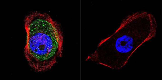
Immunofluorescent analysis of AKAP450 in A431 cells. Cells were grown on slides and fixed with formaldehyde prior to staining. Cells were probed without (control) or with AKAP450 antibody [17G10] at a dilution of 1:100 overnight at 4°C, washed with PBS and incubated with a proper secondary antibody. AKAP450 staining (green), F-Actin staining with Phalloidin (red) and nuclei with DAPI (blue) is shown. Images were taken at 60X magnification.Application
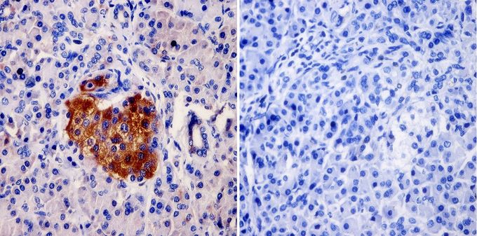
Immunohistochemistry was performed on normal biopsies of deparaffinized human pancreas tissue. To expose target proteins, heat induced antigen retrieval was performed using 10mM sodium citrate (pH6.0) buffer, microwaved for 8-15 minutes. Following antigen retrieval tissues were blocked in 3% BSA-PBS for 30 minutes at room temperature and probed at a dilution of 1:20 with or without AKAP450 antibody [17G10] overnight at 4°C in a humidified chamber. Tissues were washed with PBST and endogenous peroxidase activity was quenched with a peroxidase suppressor. Detection was performed using a biotin-conjugated secondary antibody and SA-HRP, followed by colorimetric detection using DAB. Tissues were counterstained with hematoxylin and prepped for mounting.| Product Name | AKAP450 antibody |
|---|---|
| Antibody Type | Primary Antibodies |
| Product description | Mouse monoclonal antibody [17G10] to AKAP450The A-kinase anchor proteins (AKAPs) are a group of structurally diverse proteins which have the common function of binding to the regulatory subunit of protein kinase A (PKA) and confining the holoenzyme to discrete locations within the cell. This gene encodes a member of the AKAP family. Alternate splicing of this gene results in at least two isoforms that localize to the centrosome and the Golgi apparatus, and interact with numerous signaling proteins from multiple signal transduction pathways. These signaling proteins include type II protein kinase A, serine/threonine kinase protein kinase N, protein phosphatase 1, protein phosphatase 2a, protein kinase C-epsilon and phosphodiesterase 4D3. [provided by RefSeq, Aug 2008] |
| Immunogen | Synthetic peptide corresponding to residues Q(31) F R Q R K A Q S D G Q S P S(45) of human AKAP450. |
| Clonality | Monoclonal |
|---|---|
| Isotype | IgG |
| Clone Number | 17G10 |
| Host Species | Mouse |
| Tested Applications | ICC/IFIHCIHC-PIPWB |
| IHC-P: 1:10 - 1:100: |
|
| Species Reactivity | HumanMouse |
| Concentration | 1mg/ml (Please refer to the vial label for the specific concentration) |
| Purification | Protein G |
| Gene Synonyms | YOTIAO CG-NAP PRKA9 AKAP9 PPP1R45 MU-RMS-40.16A HYPERION LQT11 A kinase (PRKA) anchor protein (yotiao) 9 AKAP350 AKAP-9 |
|---|---|
| Target | AKAP450 |
Application
Immunofluorescent analysis of AKAP450 in A431 cells. Cells were grown on slides and fixed with formaldehyde prior to staining. Cells were probed without (control) or with AKAP450 antibody [17G10] at a dilution of 1:100 overnight at 4°C, washed with PBS and incubated with a proper secondary antibody. AKAP450 staining (green), F-Actin staining with Phalloidin (red) and nuclei with DAPI (blue) is shown. Images were taken at 60X magnification.
Application
Immunohistochemistry was performed on normal biopsies of deparaffinized human pancreas tissue. To expose target proteins, heat induced antigen retrieval was performed using 10mM sodium citrate (pH6.0) buffer, microwaved for 8-15 minutes. Following antigen retrieval tissues were blocked in 3% BSA-PBS for 30 minutes at room temperature and probed at a dilution of 1:20 with or without AKAP450 antibody [17G10] overnight at 4°C in a humidified chamber. Tissues were washed with PBST and endogenous peroxidase activity was quenched with a peroxidase suppressor. Detection was performed using a biotin-conjugated secondary antibody and SA-HRP, followed by colorimetric detection using DAB. Tissues were counterstained with hematoxylin and prepped for mounting.| Application Notes | IHC-P: 1:10 - 1:100: |
|---|
| Form | Liquid |
|---|---|
| Storage Instructions | -20° C, Avoid Freeze/Thaw Cycles |
| Storage Buffer | PBS containing 0.05 % sodium azide and 1 mg/ml BSA |


