Application
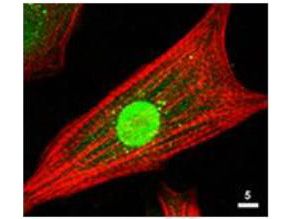
ICC/IF of the Akt antibody is used at 1: 80 for 1hr at RT on cultured neonatal rat cardiomyocytes (Green). The red is Texas-red phalloidin that labels actin filaments.Application
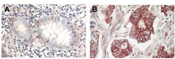
Immunohistochemical analysis of formalin-fixed paraffin-embedded tissue, A: Normal colon tissue, B: Colon tumor tissue, using AKT antibody at 1:1,000 dilution for 1 h at RT. Peroxidase rabbit secondary antibody at 1:10,000 for 45 min at RT. Localization: AKT is nuclear. Staining: AKT as precipitated red signal with hematoxylin purple nuclear counterstain.Application
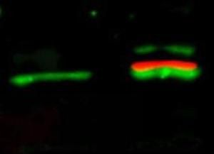
Western Blot of simultaneous detection of unphosphorylated and phosphorylated Rabbit Anti-AKT antibody . Lane 1: unstimulated NIH/3T3 lysates contain inactive unphosphorylated Akt1, green band. Lane 2: PDGF stimulated NIH/3T3 lysate contains both inactive (green band) and activated phosphorylated Akt1 (red band). Load: 35 µg per lane. Primary antibody: rabbit anti-Akt (pan) and mouse anti-Akt pS473 specific antibodies at 1:1000 for overnight at 4°C. Secondary antibody: Flourescent 549 conjugated anti-rabbit IgG (green) and flourescent 649 conjugated anti-mouse IgG (red) secondary antibodies at 1:10,000 for 45 min at RT.Application
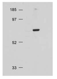
Western blot using Akt antibody at 1:500 for overnight at 4°C on 20 ug NIH/3T3 whole cell lysate.Application
Application| Product Name | AKT antibody |
|---|---|
| Antibody Type | Primary Antibodies |
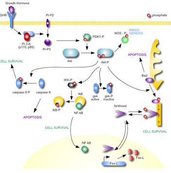
| Product description | Rabbit Polyclonal antibody to AKTAkt (Protein Kinase B, PKB) is a key regulator of many signal transduction pathways (reviewed in ref 1). Its tight control over cell proliferation and cell viability are manifold; overexpression or inappropriate activation of Akt has been seen in many types of cancer. Akt mediates many of the downstream events of PI 3-kinase (a lipid kinase activated by growth factors, cytokines and insulin). PI 3-kinase recruits Akt to the membrane, where it is activated by PDK1 phosphorylation (reviewed in 2). Once phosphorylated, Akt dissociates from the membrane and phosphorylates targets in the cytoplasm and the cell nucleus. Akt has two main roles: (i) inhibition of apoptosis; (ii) promotion of proliferation. |
| Immunogen | AKT Antibody was produced from whole rabbit serum prepared by repeated immunizations with a synthetic peptide C-R-P-H-F-P-Q-F-S-Y-S-A-S-G-T-A corresponding to the C-terminus (460-480) of human, rat and mouse and chicken AKT proteins conjugated to KLH usin |
| Clonality | Polyclonal |
|---|---|
| Host Species | Rabbit |
| Tested Applications | ELISAFACSICC/IFIHC-PWB |
| *Optimal dilutions/concentrations should be determined by the researcher. | |
| Species Reactivity | HumanMouseChickenRat |
| Concentration | 1mg/ml |
| Purification | Affinity purified |
| Target | Akt |
|---|
Application
ICC/IF of the Akt antibody is used at 1: 80 for 1hr at RT on cultured neonatal rat cardiomyocytes (Green). The red is Texas-red phalloidin that labels actin filaments.
Application
Immunohistochemical analysis of formalin-fixed paraffin-embedded tissue, A: Normal colon tissue, B: Colon tumor tissue, using AKT antibody at 1:1,000 dilution for 1 h at RT. Peroxidase rabbit secondary antibody at 1:10,000 for 45 min at RT. Localization: AKT is nuclear. Staining: AKT as precipitated red signal with hematoxylin purple nuclear counterstain.
Application
Western Blot of simultaneous detection of unphosphorylated and phosphorylated Rabbit Anti-AKT antibody . Lane 1: unstimulated NIH/3T3 lysates contain inactive unphosphorylated Akt1, green band. Lane 2: PDGF stimulated NIH/3T3 lysate contains both inactive (green band) and activated phosphorylated Akt1 (red band). Load: 35 µg per lane. Primary antibody: rabbit anti-Akt (pan) and mouse anti-Akt pS473 specific antibodies at 1:1000 for overnight at 4°C. Secondary antibody: Flourescent 549 conjugated anti-rabbit IgG (green) and flourescent 649 conjugated anti-mouse IgG (red) secondary antibodies at 1:10,000 for 45 min at RT.
Application
Western blot using Akt antibody at 1:500 for overnight at 4°C on 20 ug NIH/3T3 whole cell lysate.
Application
Application| Application Notes | *Optimal dilutions/concentrations should be determined by the researcher. |
|---|
| Form | Liquid |
|---|---|
| Storage Instructions | Store vial at -20° C prior to opening. Aliquot contents and freeze at -20° C or below for extended storage. Avoid cycles of freezing and thawing. |
| Storage Buffer | 0.02 M Potassium Phosphate, 0.15 M Sodium Chloride, pH 7.2 and 0.01% (w/v) Sodium Azide |


