Application
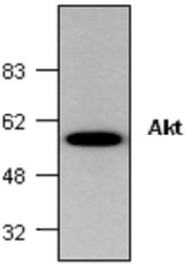
Western blot analysis of Akt expression in mouse small intestine tissue lysate.WB
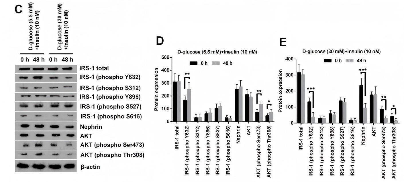
(C–E) The phosphorylation of insulin receptor substrate (IRS)-1 (phospho Y632), AKT (phospho Ser473) and AKT (phospho Thr308) was markedly reduced by HG inducement.WB
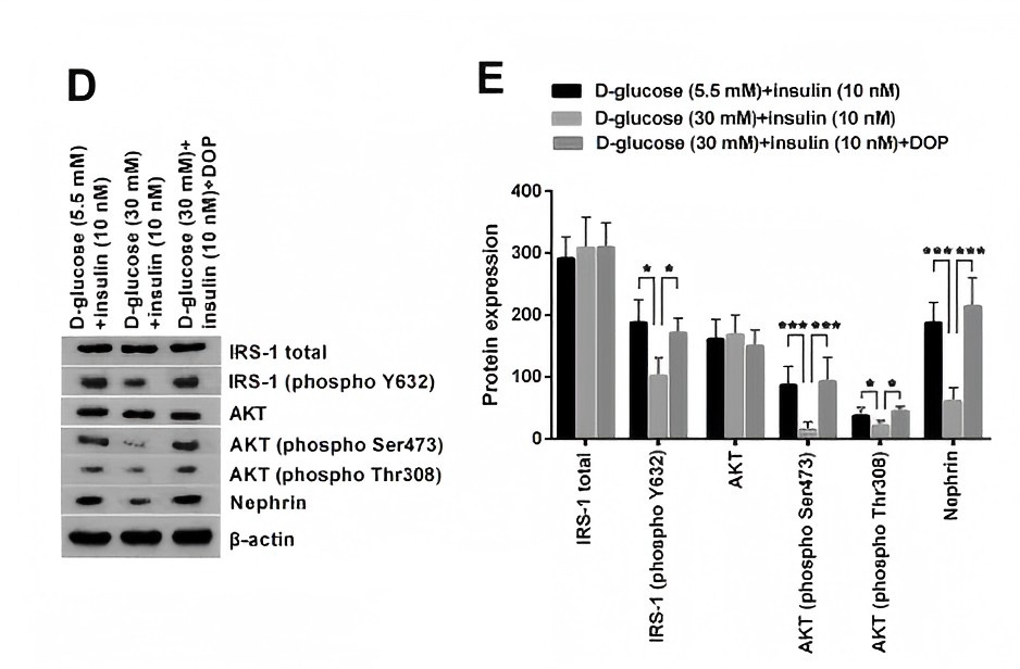
(D, E) DOP treatment markedly relived HG-induced suppressive effects on the phosphorylation of insulin receptor substrate (IRS)-1 (phospho Y632), AKT (phospho Ser473), AKT (phospho Thr308) and Nephrin.WB
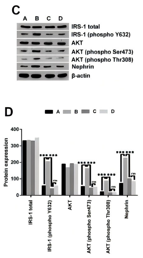
(C, D) IRS-1/2 inhibitor NT157 treatment notably reversed HG-induced increased phosphorylation of IRS-1 (phospho Y632), AKT (phospho Ser473), AKT (phospho Thr308) and Nephrin.WB
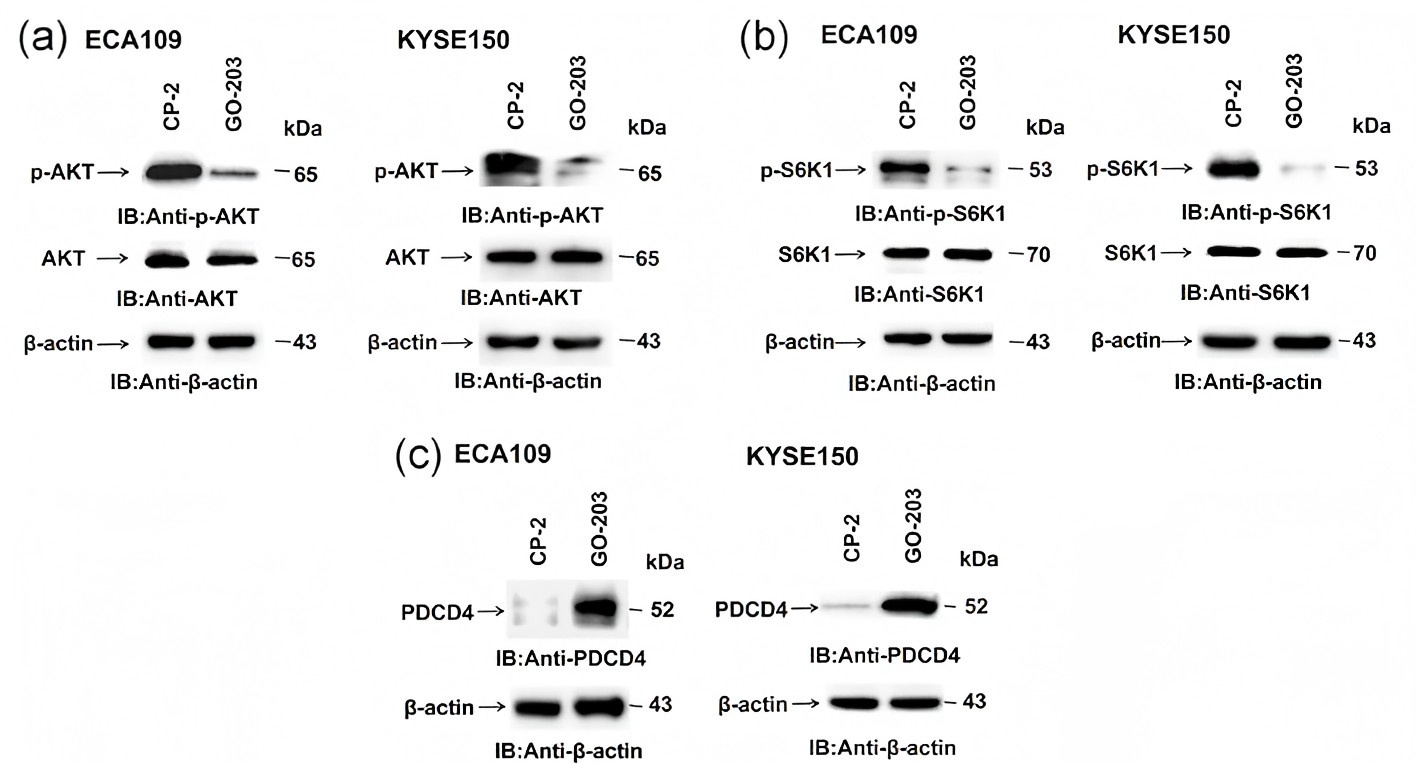
(a–c) ECA109 and KYSE150 cells were treated each day with 5 μM GO-203 or CP-2 for 3 days, and GO-203 inhibits the phosphorylation of AKT and S6K1 protein and then increases PDCD4 leave in ESCC cells.WB
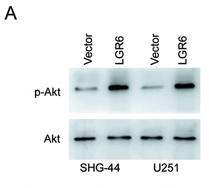
Following LGR6 overexpression, protein expression levels were (A) determined by western blotting.WB
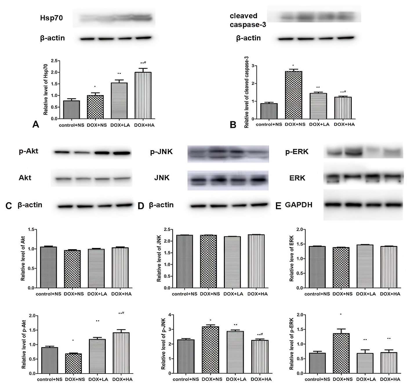
A–E, Western blotting evaluations of Hsp70 (A), cleaved caspase-3 (B), p-Akt and Akt (C), p-JNK and JNK (D), and p-ERK and ERK (E) levels in the myocardium.| Product Name | AKT antibody |
|---|---|
| Antibody Type | Primary Antibodies |
| Product description | Rabbit polyclonal antibody to AktAkt, also referred to as PKB and RAC, is the cellular homologue of the viral oncogene v-Akt. This protein kinase is activated by insulin and various growth factors and functions in a pathway involving PI3 kinase as activation is sensitive to wortmanin. Recent evidence suggests that Akt functions to promote cell survival by actively inhibiting apoptosis. |
| Clonality | Polyclonal |
|---|---|
| Isotype | IgG |
| Host Species | Rabbit |
| Tested Applications | IHCIPWB |
| WB:1:250~1:1000 IP:1:50~1:250 IHC:1:25~1:100 |
|
| Species Reactivity | BovineHumanMouseRat |
| Concentration | 0.5mg/ml |
| Purification | Affinity purified |
| Gene Synonyms | Akt1, RAC, Akt 2, Akt2, Akt 1, Akt3, Akt 3, PKB |
|---|---|
| Target | Akt |
| Tissue Specificity | This antibody is specific for Akt/PKB. |
Application
Western blot analysis of Akt expression in mouse small intestine tissue lysate.
WB
(C–E) The phosphorylation of insulin receptor substrate (IRS)-1 (phospho Y632), AKT (phospho Ser473) and AKT (phospho Thr308) was markedly reduced by HG inducement.
WB
(D, E) DOP treatment markedly relived HG-induced suppressive effects on the phosphorylation of insulin receptor substrate (IRS)-1 (phospho Y632), AKT (phospho Ser473), AKT (phospho Thr308) and Nephrin.
WB
(C, D) IRS-1/2 inhibitor NT157 treatment notably reversed HG-induced increased phosphorylation of IRS-1 (phospho Y632), AKT (phospho Ser473), AKT (phospho Thr308) and Nephrin.
WB
(a–c) ECA109 and KYSE150 cells were treated each day with 5 μM GO-203 or CP-2 for 3 days, and GO-203 inhibits the phosphorylation of AKT and S6K1 protein and then increases PDCD4 leave in ESCC cells.
WB
Following LGR6 overexpression, protein expression levels were (A) determined by western blotting.
WB
A–E, Western blotting evaluations of Hsp70 (A), cleaved caspase-3 (B), p-Akt and Akt (C), p-JNK and JNK (D), and p-ERK and ERK (E) levels in the myocardium.| Application Notes | WB:1:250~1:1000 IP:1:50~1:250 IHC:1:25~1:100 |
|---|
| Form | Liquid |
|---|---|
| Storage Instructions | The antibody solution should be gently mixed before use.Store at -20 °C. |
| Storage Buffer | PBS (pH 7.2) containing 30% glycerol, 0.5% BSA, 0.01% thimerosal |


