Application
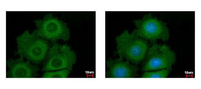
Akt1/2/3 antibody detects Akt1/2/3 protein at cytoplasm by immunofluorescent analysis. Sample: MCF-7 cells were fixed in ice-cold MeOH for 5 min. Green: Akt1 protein stained by Akt1/2/3 antibody diluted at 1:500. Blue: Hoechst 33342 staining.Application
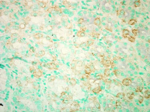
Staining of Human normal esophagus tissue sections using anti-Akt1/2/3 antibody .Application
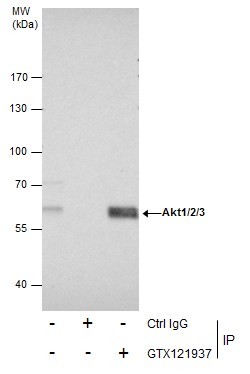
Immunoprecipitation of Akt1/2/3 protein from 293T whole cell extracts using 5 μg of Akt1/2/3 antibody [N3C2], Internal . Western blot analysis was performed using Akt1/2/3 antibody [N3C2], Internal . EasyBlot anti-Rabbit IgG was used as a secondary reagent.Application
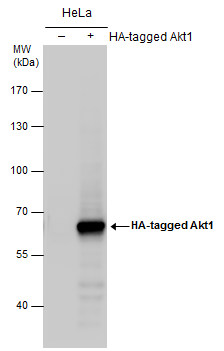
Non-transfected (–) and transfected (+) HeLa whole cell extracts (30 µg) were separated by 7.5% SDS-PAGE, and the membrane was blotted with AKT antibody [N3C2], Internal diluted at 1:5000. The HRP-conjugated anti-rabbit IgG antibody was used to detect the primary antibody.Application
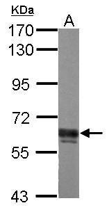
Sample (30 µg of whole cell lysate) A: PC-12 7.5% SDS PAGE diluted at 1:3000 The HRP-conjugated anti-rabbit IgG antibody was used to detect the primary antibody.Application
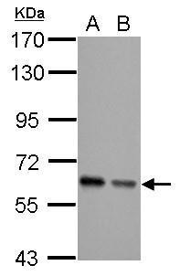
Sample (30 µg of whole cell lysate) A: JC B: BCL-1 7.5% SDS PAGE diluted at 1:5000 The HRP-conjugated anti-rabbit IgG antibody was used to detect the primary antibody.Application
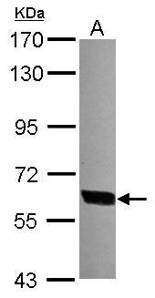
Sample (30 µg of whole cell lysate) A: 293T 7.5% SDS PAGE diluted at 1:10000 The HRP-conjugated anti-rabbit IgG antibody was used to detect the primary antibody.| Product Name | AKT antibody , Internal |
|---|---|
| Antibody Type | Primary Antibodies |
| Product description | Rabbit Polyclonal antibody to AKT1+2+3 (v-akt murine thymoma viral oncogene homolog 1)The serine-threonine protein kinase encoded by the AKT1 gene is catalytically inactive in serum-starved primary and immortalized fibroblasts. AKT1 and the related AKT2 are activated by platelet-derived growth factor. The activation is rapid and specific, and it is abrogated by mutations in the pleckstrin homology domain of AKT1. It was shown that the activation occurs through phosphatidylinositol 3-kinase. In the developing nervous system AKT is a critical mediator of growth factor-induced neuronal survival. Survival factors can suppress apoptosis in a transcription-independent manner by activating the serine/threonine kinase AKT1, which then phosphorylates and inactivates components of the apoptotic machinery. Multiple alternatively spliced transcript variants have been found for this gene. [provided by RefSeq] |
| Immunogen | Recombinant protein encompassing a sequence within the center region of human Akt1/2/3. The exact sequence is proprietary. |
| Clonality | Polyclonal |
|---|---|
| Isotype | IgG |
| Host Species | Rabbit |
| Tested Applications | ICC/IFIHCIHC-PIPWB |
| *Optimal dilutions/concentrations should be determined by the researcher. | |
| Species Reactivity | HumanMouseRat |
| Predicted Reactivity | Zebrafish, Xenopus laevis, Cat, Pig, Chicken, Sheep, Rhesus Monkey, BovineZebrafish (93%), Xenopus laevis (99%), Cat (98%), Pig (99%), Chicken (100%), Sheep (99%), Rhesus Monkey (100%), Bovine (99%) |
| Concentration | 1mg/ml (Please refer to the vial label for the specific concentration) |
| Purification | Affinity purified |
| Gene Synonyms | AKT antibody, MGC99656 antibody, PKB antibody, PKB-ALPHA antibody, PRKBA antibody, RAC antibody, RAC-ALPHA antibody, AKT1 antibody, protein kinase B alpha antibody, rac protein kinase alpha antibody, RAC-alpha serine/threonine-protein kinase antibody, prot |
|---|---|
| Molecular Weight(MW) | 56 kDa (note) (4) |
| Tissue Specificity | Knockdown/Knockout validation was supported by customer review data. |
| Cellular Localization | Cytoplasm , Nucleus , Cell membrane |
Application
Akt1/2/3 antibody detects Akt1/2/3 protein at cytoplasm by immunofluorescent analysis. Sample: MCF-7 cells were fixed in ice-cold MeOH for 5 min. Green: Akt1 protein stained by Akt1/2/3 antibody diluted at 1:500. Blue: Hoechst 33342 staining.
Application
Staining of Human normal esophagus tissue sections using anti-Akt1/2/3 antibody .
Application
Immunoprecipitation of Akt1/2/3 protein from 293T whole cell extracts using 5 μg of Akt1/2/3 antibody [N3C2], Internal . Western blot analysis was performed using Akt1/2/3 antibody [N3C2], Internal . EasyBlot anti-Rabbit IgG was used as a secondary reagent.
Application
Non-transfected (–) and transfected (+) HeLa whole cell extracts (30 µg) were separated by 7.5% SDS-PAGE, and the membrane was blotted with AKT antibody [N3C2], Internal diluted at 1:5000. The HRP-conjugated anti-rabbit IgG antibody was used to detect the primary antibody.
Application
Sample (30 µg of whole cell lysate) A: PC-12 7.5% SDS PAGE diluted at 1:3000 The HRP-conjugated anti-rabbit IgG antibody was used to detect the primary antibody.
Application
Sample (30 µg of whole cell lysate) A: JC B: BCL-1 7.5% SDS PAGE diluted at 1:5000 The HRP-conjugated anti-rabbit IgG antibody was used to detect the primary antibody.
Application
Sample (30 µg of whole cell lysate) A: 293T 7.5% SDS PAGE diluted at 1:10000 The HRP-conjugated anti-rabbit IgG antibody was used to detect the primary antibody.| Application Notes | *Optimal dilutions/concentrations should be determined by the researcher. |
|---|
| Form | Liquid |
|---|---|
| Storage Instructions | Keep as concentrated solution. Aliquot and store at -20ºC or below. Avoid multiple freeze-thaw cycles. |
| Storage Buffer | 1XPBS, 1% BSA, 20% Glycerol (pH7). 0.01% Thimerosal was added as a preservative. |


