Application
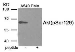
Western blot analysis of extracts from A549 cells treated with PMA using Phospho-Akt (Ser129) antibody. The lane on the right is treated with the antigen-specific peptide.| Product Name | AKT1 (phospho Ser129) antibody |
|---|---|
| Antibody Type | Primary Antibodies |
| Product description | Rabbit Polyclonal antibody to AKT1 (phospho Ser129)The serine-threonine protein kinase encoded by the AKT1 gene is catalytically inactive in serum-starved primary and immortalized fibroblasts. AKT1 and the related AKT2 are activated by platelet-derived growth factor. The activation is rapid and specific, and it is abrogated by mutations in the pleckstrin homology domain of AKT1. It was shown that the activation occurs through phosphatidylinositol 3-kinase. In the developing nervous system AKT is a critical mediator of growth factor-induced neuronal survival. Survival factors can suppress apoptosis in a transcription-independent manner by activating the serine/threonine kinase AKT1, which then phosphorylates and inactivates components of the apoptotic machinery. Mutations in this gene have been associated with the Proteus syndrome. Multiple alternatively spliced transcript variants have been found for this gene. [provided by RefSeq, Jul 2011] |
| Immunogen | Peptide sequence around phosphorylation site of serine129(D-N-S(p)-G-A) derived from Human Akt . |
| Modification | p-Ser129 |
| Clonality | Polyclonal |
|---|---|
| Isotype | IgG |
| Host Species | Rabbit |
| Tested Applications | WB |
| WB: 1:500 - 1:1000 | |
| Species Reactivity | HumanMouseRat |
| Concentration | 1mg/ml (Please refer to the vial label for the specific concentration) |
| Purification | Affinity purified |
| Gene Synonyms | v-akt murine thymoma viral oncogene homolog 1 , PKB , PRKBA , AKT1 , CWS6 , RAC-ALPHA , AKT , RAC , PKB-ALPHA |
|---|---|
| Target | AKT1 (phospho Ser129) |
| Tissue Specificity | The antibody detects endogenous level of Akt only when phosphorylated at serine 129. |
Application
Western blot analysis of extracts from A549 cells treated with PMA using Phospho-Akt (Ser129) antibody. The lane on the right is treated with the antigen-specific peptide.| Application Notes | WB: 1:500 - 1:1000 |
|---|
| Form | Liquid |
|---|---|
| Storage Instructions | Store at -20˚C/1 year |
| Storage Buffer | Phosphate buffered saline (without Mg2+ and Ca2+), pH 7.4, 150mM NaCl, 0.02% sodium azide and 50% glycerol. |


