Application
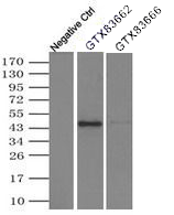
IP analysis was performed with 500ul of DDK tagged SERPINA1 overexpression lysates and either no antibody control, 2ug of or 2ug of The precipitated protein was detected by DDK tag antibody.Application
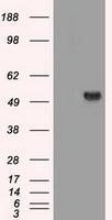
HEK293T cells were transfected with the pCMV6-ENTRY control (Left lane) or pCMV6-ENTRY SERPINA1 (Right lane) cDNA for 48 hrs and lysed. Equivalent amounts of cell lysates (5 ug per lane) were separated by SDS-PAGE and immunoblotted with anti-SERPINA1.Application
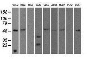
Western blot analysis of extracts (35ug) from 9 different cell lines by using anti-SERPINA1 monoclonal antibody.Application
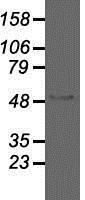
Western blot analysis of 35ug of cell extracts from human brain tissue lysates using anti-SERPINA1 antibody.Application
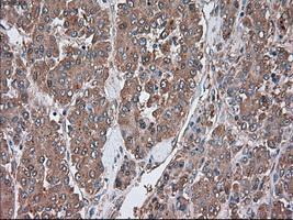
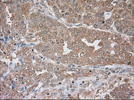
Immunohistochemical staining of paraffin-embedded Carcinoma of liver tissue using anti-SERPINA1mouse monoclonal antibody. ( Dilution 1:50)Application
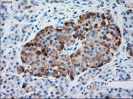
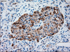
Immunohistochemical staining of paraffin-embedded pancreas tissue using anti-SERPINA1mouse monoclonal antibody. ( Dilution 1:50)Application
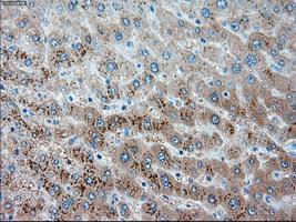
Immunohistochemical staining of paraffin-embedded liver tissue using anti-SERPINA1mouse monoclonal antibody. ( Dilution 1:50)Application
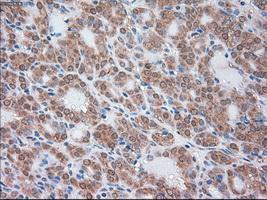
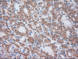
Immunohistochemical staining of paraffin-embedded Carcinoma of thyroid tissue using anti-SERPINA1mouse monoclonal antibody. ( Dilution 1:50)Application
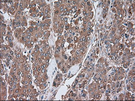
Immunohistochemical staining of paraffin-embedded Adenocarcinoma of endometrium tissue using anti-SERPINA1mouse monoclonal antibody. ( Dilution 1:50)Application
Immunohistochemical staining of paraffin-embedded carcinoma of human liver tissue using anti-SERPINA1 mouse monoclonal antibody.Application
Immunohistochemical staining of paraffin-embedded carcinoma of human thyroid tissue using anti-SERPINA1 mouse monoclonal antibody.Application
Immunohistochemical staining of paraffin-embedded human pancreas tissue using anti-SERPINA1 mouse monoclonal antibody.Application
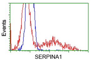
HEK293T cells transfected with either SERPINA1 overexpress plasmid (Red) or empty vector control plasmid (Blue) were immunostained by anti-SERPINA1 antibody , and then analyzed by flow cytometry.| Product Name | alpha 1 Antitrypsin antibody |
|---|---|
| Antibody Type | Primary Antibodies |
| Product description | Mouse monoclonal antibody [9A1] to alpha 1 AntitrypsinInhibitor of serine proteases. Its primary target is elastase, but it also has a moderate affinity for plasmin and thrombin. Irreversibly inhibits trypsin, chymotrypsin and plasminogen activator. The aberrant form inhibits insulin-induced NO synthesis in platelets, decreases coagulation time and has proteolytic activity against insulin and plasmin. Short peptide from AAT (SPAAT) is a reversible chymotrypsin inhibitor. It also inhibits elastase, but not trypsin. Its major physiological function is the protection of the lower respiratory tract against proteolytic destruction by human leukocyte elastase (HLE). |
| Immunogen | Protein expressed in 293T cell transfected with human SERPINA1 expression vector |
| Clonality | Monoclonal |
|---|---|
| Isotype | IgG1 |
| Clone Number | 9A1 |
| Host Species | Mouse |
| Tested Applications | FACSIHC-PIPWB |
| ICC/IF (1:100): |
|
| Species Reactivity | HumanDogMonkey |
| Concentration | 1mg/ml |
| Purification | Affinity purified |
| Gene Synonyms | AAT PI1 MGC23330 PRO2275 SERPINA1 A1AT MGC9222 PI A1A |
|---|---|
| Target | alpha 1 Antitrypsin |
| Tissue Specificity | is specific for human SERPINA1. |
Application
IP analysis was performed with 500ul of DDK tagged SERPINA1 overexpression lysates and either no antibody control, 2ug of or 2ug of The precipitated protein was detected by DDK tag antibody.
Application
HEK293T cells were transfected with the pCMV6-ENTRY control (Left lane) or pCMV6-ENTRY SERPINA1 (Right lane) cDNA for 48 hrs and lysed. Equivalent amounts of cell lysates (5 ug per lane) were separated by SDS-PAGE and immunoblotted with anti-SERPINA1.
Application
Western blot analysis of extracts (35ug) from 9 different cell lines by using anti-SERPINA1 monoclonal antibody.
Application
Western blot analysis of 35ug of cell extracts from human brain tissue lysates using anti-SERPINA1 antibody.
Application
Immunohistochemical staining of paraffin-embedded Carcinoma of liver tissue using anti-SERPINA1mouse monoclonal antibody. ( Dilution 1:50)
Application
Immunohistochemical staining of paraffin-embedded pancreas tissue using anti-SERPINA1mouse monoclonal antibody. ( Dilution 1:50)
Application
Immunohistochemical staining of paraffin-embedded liver tissue using anti-SERPINA1mouse monoclonal antibody. ( Dilution 1:50)
Application
Immunohistochemical staining of paraffin-embedded Carcinoma of thyroid tissue using anti-SERPINA1mouse monoclonal antibody. ( Dilution 1:50)
Application
Immunohistochemical staining of paraffin-embedded Adenocarcinoma of endometrium tissue using anti-SERPINA1mouse monoclonal antibody. ( Dilution 1:50)
Application
Immunohistochemical staining of paraffin-embedded carcinoma of human liver tissue using anti-SERPINA1 mouse monoclonal antibody.
Application
Immunohistochemical staining of paraffin-embedded carcinoma of human thyroid tissue using anti-SERPINA1 mouse monoclonal antibody.
Application
Immunohistochemical staining of paraffin-embedded human pancreas tissue using anti-SERPINA1 mouse monoclonal antibody.
Application
HEK293T cells transfected with either SERPINA1 overexpress plasmid (Red) or empty vector control plasmid (Blue) were immunostained by anti-SERPINA1 antibody , and then analyzed by flow cytometry.| Application Notes | ICC/IF (1:100): |
|---|
| Form | Liquid |
|---|---|
| Storage Instructions | For short-term storage, store at 4°C or aliquot into working amounts and store at -20°C. For long-term storage, store at -70°C (aliquotted). Avoid repeated freeze-thaw cycles. |
| Storage Buffer | PBS (PH 7.3) containing 1% BSA, 50% glycerol and 0.02% sodium azide. |


