Application
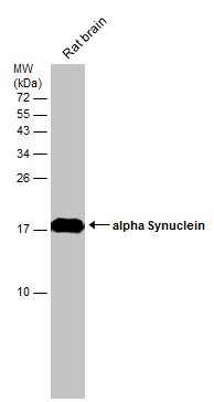
Rat tissue extract (50 µg) was separated by 15% SDS-PAGE, and the membrane was blotted with alpha Synuclein antibody diluted at 1:1000.Application
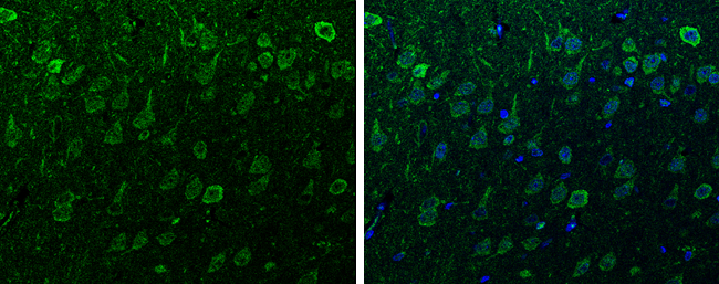
alpha Synuclein antibody detects alpha Synuclein protein at cytoplasm and nucleus in rat brain by immunohistochemical analysis. Sample: Paraffin-embedded rat brain. Green: alpha Synuclein antibody diluted at 1:200. The signal was developed using goat anti-rabbit IgG antibody (Dylight488) . Blue: Nuclear staining with Hoechst 33342.Application
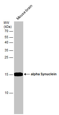
Mouse tissue extract (50 µg) was separated by 15% SDS-PAGE, and the membrane was blotted with alpha Synuclein antibody diluted at 1:1000.Application
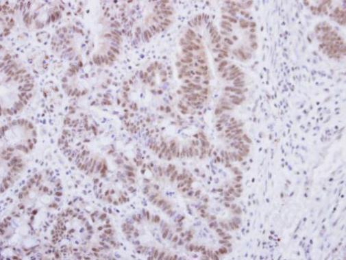
Immunohistochemical analysis of paraffin-embedded human colon carcinoma, using alpha Synuclein antibody at 1:250 dilution.Application
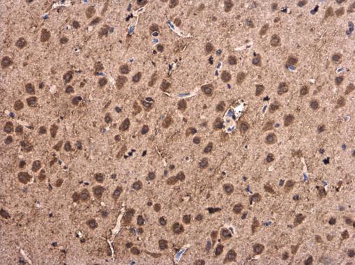
alpha Synuclein antibody detects alpha Synuclein protein at cytoplasm and nucleus in Rat brain by immunohistochemical analysis. Sample: Paraffin-embedded Rat brain. alpha Synuclein antibody diluted at 1:500.Application
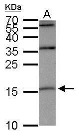
alpha Synuclein antibody detects SNCA protein by western blot analysis. A. 30 µg SK-N-AS whole cell lysate/extract 15% SDS-PAGE alpha Synuclein antibody dilution: 1:500 The HRP-conjugated anti-rabbit IgG antibody was used to detect the primary antibody.Application
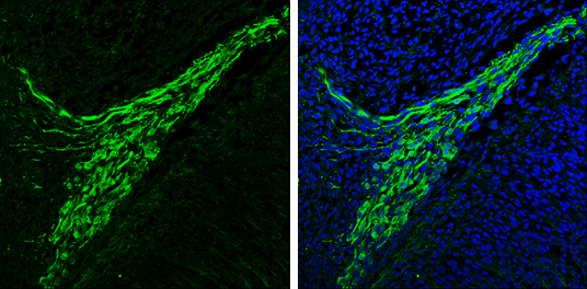
alpha Synuclein antibody detects alpha Synuclein protein expression by immunohistochemical analysis. Sample: Frozen sectioned E13.5 Rat brain. Green: alpha Synuclein protein stained by alpha Synuclein antibody diluted at 1:250. Blue: Fluoroshield with DAPI .Application
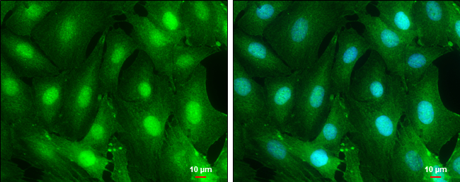
alpha Synuclein antibody detects alpha Synuclein protein at cytoplasm and nucleus by immunofluorescent analysis. Sample: SK-N-SH cells were fixed in 4% paraformaldehyde at RT for 15 min. Green: alpha Synuclein protein stained by alpha Synuclein antibody diluted at 1:500. Blue: Hoechst 33342 staining. Scale bar = 10 μm.| Product Name | alpha Synuclein antibody |
|---|---|
| Antibody Type | Primary Antibodies |
| Product description | Rabbit Polyclonal antibody to alpha Synuclein (synuclein, alpha (non A4 component of amyloid precursor))Alpha-synuclein is a member of the synuclein family, which also includes beta- and gamma-synuclein. Synucleins are abundantly expressed in the brain and alpha- and beta-synuclein inhibit phospholipase D2 selectively. SNCA may serve to integrate presynaptic signaling and membrane trafficking. Defects in SNCA have been implicated in the pathogenesis of Parkinson disease. SNCA peptides are a major component of amyloid plaques in the brains of patients with Alzheimer's disease. Four alternatively spliced transcripts encoding two different isoforms have been identified for this gene. [provided by RefSeq] |
| Immunogen | Full length human alpha Synuclein Recombinant protein. |
| Clonality | Polyclonal |
|---|---|
| Isotype | IgG |
| Host Species | Rabbit |
| Tested Applications | ICC/IFIHC-FIHC-PWB |
| *Optimal dilutions/concentrations should be determined by the researcher.
: |
|
| Species Reactivity | HumanMouseRat |
| Predicted Reactivity | Xenopus laevis, Pig, Chicken, Sheep, Chimpanzee, BovineXenopus laevis (85%), Pig (97%), Chicken (88%), Sheep (95%), Chimpanzee (100%), Bovine (94%) |
| Concentration | 1mg/ml |
| Purification | Affinity purified |
| Gene Synonyms | MGC110988 antibody NACP antibody PARK1 antibody PARK4 antibody PD1 antibody SNCA antibody non A-beta component of AD amyloid antibody alpha-synuclein antibody alpha-synuclein isoform NACP140 antibody synuclein alpha-140 antibody synuclein alpha |
|---|---|
| Molecular Weight(MW) | 14 kDa (note) (4) |
| Cellular Localization | Cytoplasm , Membrane , Nucleus |
Application
Rat tissue extract (50 µg) was separated by 15% SDS-PAGE, and the membrane was blotted with alpha Synuclein antibody diluted at 1:1000.
Application
alpha Synuclein antibody detects alpha Synuclein protein at cytoplasm and nucleus in rat brain by immunohistochemical analysis. Sample: Paraffin-embedded rat brain. Green: alpha Synuclein antibody diluted at 1:200. The signal was developed using goat anti-rabbit IgG antibody (Dylight488) . Blue: Nuclear staining with Hoechst 33342.
Application
Mouse tissue extract (50 µg) was separated by 15% SDS-PAGE, and the membrane was blotted with alpha Synuclein antibody diluted at 1:1000.
Application
Immunohistochemical analysis of paraffin-embedded human colon carcinoma, using alpha Synuclein antibody at 1:250 dilution.
Application
alpha Synuclein antibody detects alpha Synuclein protein at cytoplasm and nucleus in Rat brain by immunohistochemical analysis. Sample: Paraffin-embedded Rat brain. alpha Synuclein antibody diluted at 1:500.
Application
alpha Synuclein antibody detects SNCA protein by western blot analysis. A. 30 µg SK-N-AS whole cell lysate/extract 15% SDS-PAGE alpha Synuclein antibody dilution: 1:500 The HRP-conjugated anti-rabbit IgG antibody was used to detect the primary antibody.
Application
alpha Synuclein antibody detects alpha Synuclein protein expression by immunohistochemical analysis. Sample: Frozen sectioned E13.5 Rat brain. Green: alpha Synuclein protein stained by alpha Synuclein antibody diluted at 1:250. Blue: Fluoroshield with DAPI .
Application
alpha Synuclein antibody detects alpha Synuclein protein at cytoplasm and nucleus by immunofluorescent analysis. Sample: SK-N-SH cells were fixed in 4% paraformaldehyde at RT for 15 min. Green: alpha Synuclein protein stained by alpha Synuclein antibody diluted at 1:500. Blue: Hoechst 33342 staining. Scale bar = 10 μm.| Positive Control | SK-N-AS , mouse brain , rat brain |
|---|---|
| Application Notes | *Optimal dilutions/concentrations should be determined by the researcher.
: |
| Form | Liquid |
|---|---|
| Storage Instructions | Keep as concentrated solution. Aliquot and store at -20ºC or below. Avoid multiple freeze-thaw cycles. |
| Storage Buffer | 0.1M Tris, 0.1M Glycine, 10% Glycerol (pH7). 0.01% Thimerosal was added as a preservative. |


