Application
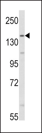
Western blot analysis of AOX1 Antibody (Center) in HepG2 cell line lysates (35ug/lane). AOX1 (arrow) was detected using the purified Pab.Application
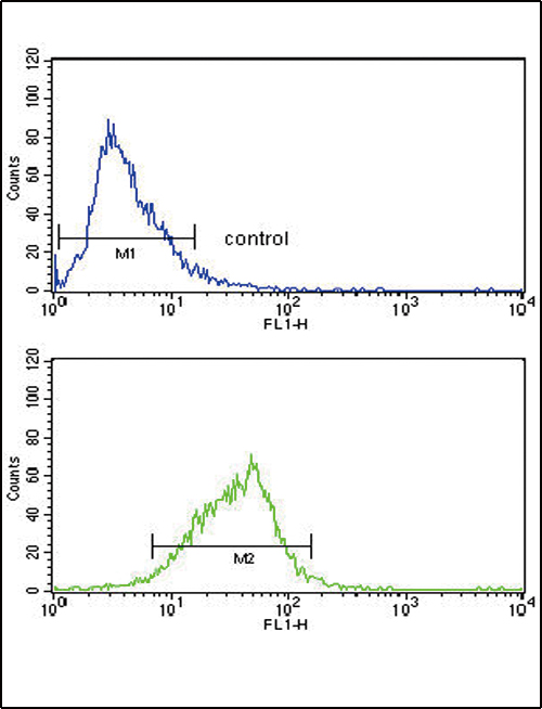
AOX1 Antibody (Center) flow cytometry analysis of HepG2 cells (bottom histogram) compared to a negative control cell (top histogram). FITC-conjugated goat-anti-rabbit secondary antibodies were used for the analysis.| Product Name | AOX1 antibody, Internal |
|---|---|
| Antibody Type | Primary Antibodies |
| Product description | Rabbit polyclonal to AOX1 (Internal) |
| Immunogen | KLH conjugated synthetic peptide between 705~734 amino acids from the Center region of human AOX1. |
| Clonality | Polyclonal |
|---|---|
| Isotype | IgG |
| Host Species | Rabbit |
| Tested Applications | FACSWB |
| ELISA: Use at 1:1,000 | |
| Species Reactivity | Human |
| Concentration | 0.2mg/ml |
| Purification | Protein G |
| Gene Synonyms | AO, AOX1, Aldehyde oxidase |
|---|---|
| Target | AOX1 |
| Molecular Weight(MW) | 147.918 kDa (note) |
Application
Western blot analysis of AOX1 Antibody (Center) in HepG2 cell line lysates (35ug/lane). AOX1 (arrow) was detected using the purified Pab.
Application
AOX1 Antibody (Center) flow cytometry analysis of HepG2 cells (bottom histogram) compared to a negative control cell (top histogram). FITC-conjugated goat-anti-rabbit secondary antibodies were used for the analysis.| Application Notes | ELISA: Use at 1:1,000 |
|---|
| Form | Liquid |
|---|---|
| Storage Instructions | Store at at 2-8°C for up to 6 months. For long term storage, aliquot and store at -20 °C. Avoid repeated freeze/thaw cycles. |
| Storage Buffer | PBS with 0.09% (W/V) sodium azide |


