Application
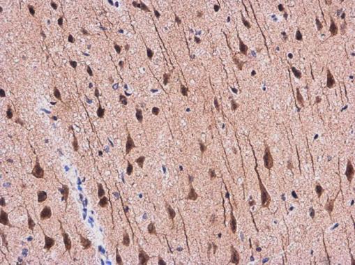
APP antibody detects APP protein at cytoplasm in Rat brain by immunohistochemical analysis. Sample: Paraffin-embedded Rat brain. APP antibody diluted at 1:500.Application
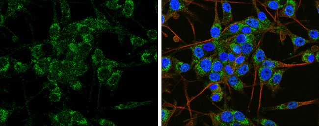
APP antibody detects APP protein at cytoplasm by immunofluorescent analysis. Sample: SH-SY5Y cells were fixed in 4% paraformaldehyde at RT for 15 min. Green: APP protein stained by APP antibody diluted at 1:100. Red: beta Tubulin 3/ TUJ1 protein stained by beta Tubulin 3/ TUJ1 antibody diluted at 1:200. Blue: Hoechst 33342 staining.Application
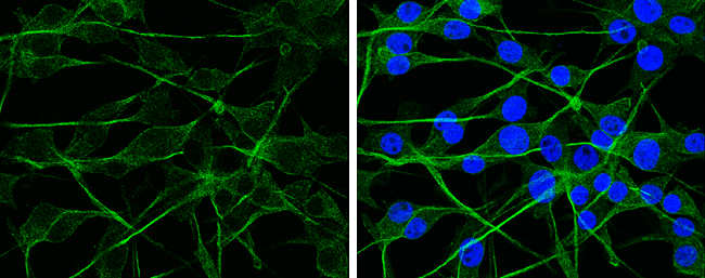
APP antibody detects APP protein at cytoplasm by immunofluorescent analysis. Sample: U-87 MG cells were fixed in 4% paraformaldehyde at RT for 15 min. Green: APP protein stained by APP antibody diluted at 1:100. Blue: Hoechst 33342 staining.Application
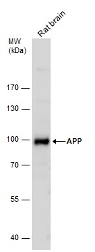
Rat tissue extract (50 μg) was separated by 7.5% SDS-PAGE, and the membrane was blotted with APP antibody diluted at 1:500.Application
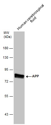
Human tissue extract (30 μg) was separated by 7.5% SDS-PAGE, and the membrane was blotted with APP antibody diluted at 1:500.Application
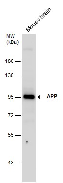
Mouse tissue extract (50 μg) was separated by 7.5% SDS-PAGE, and the membrane was blotted with APP antibody diluted at 1:500.Application
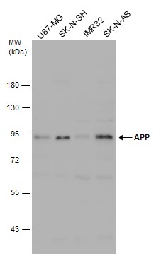
Various whole cell extracts (30 μg) were separated by 7.5% SDS-PAGE, and the membrane was blotted with APP antibody diluted at 1:500.| Product Name | APP antibody |
|---|---|
| Antibody Type | Primary Antibodies |
| Product description | Rabbit polyclonal antibody to APPThis gene encodes a cell surface receptor and transmembrane precursor protein that is cleaved by secretases to form a number of peptides. Some of these peptides are secreted and can bind to the acetyltransferase complex APBB1/TIP60 to promote transcriptional activation, while others form the protein basis of the amyloid plaques found in the brains of patients with Alzheimer disease. Mutations in this gene have been implicated in autosomal dominant Alzheimer disease and cerebroarterial amyloidosis (cerebral amyloid angiopathy). Multiple transcript variants encoding several different isoforms have been found for this gene. [provided by RefSeq] |
| Immunogen | Recombinant protein encompassing a sequence within the N-terminus region of human APP. The exact sequence is proprietary. |
| Clonality | Polyclonal |
|---|---|
| Isotype | IgG |
| Host Species | Rabbit |
| Tested Applications | ICC/IFIHC-PWB |
| *Optimal dilutions/concentrations should be determined by the researcher. | |
| Species Reactivity | HumanMouseRat |
| Predicted Reactivity | Pig, Chicken, Chimpanzee, BovinePig (81%), Chicken (80%), Chimpanzee (100%), Bovine (83%) |
| Concentration | 1mg/ml |
| Purification | Affinity purified |
| Gene Synonyms | AAA antibody, ABETA antibody, ABPP antibody, AD1 antibody, APPI antibody, CTFgamma antibody, CVAP antibody, PN2 antibody, APP antibody, alzheimer disease amyloid protein antibody, beta-amyloid peptide antibody, amyloid beta A4 protein antibody, |
|---|---|
| Target | amyloid beta A4 |
| Molecular Weight(MW) | 87 kDa (note) (7) |
| Cellular Localization | Membrane , Membrane , clathrin-coated pit |
Application
APP antibody detects APP protein at cytoplasm in Rat brain by immunohistochemical analysis. Sample: Paraffin-embedded Rat brain. APP antibody diluted at 1:500.
Application
APP antibody detects APP protein at cytoplasm by immunofluorescent analysis. Sample: SH-SY5Y cells were fixed in 4% paraformaldehyde at RT for 15 min. Green: APP protein stained by APP antibody diluted at 1:100. Red: beta Tubulin 3/ TUJ1 protein stained by beta Tubulin 3/ TUJ1 antibody diluted at 1:200. Blue: Hoechst 33342 staining.
Application
APP antibody detects APP protein at cytoplasm by immunofluorescent analysis. Sample: U-87 MG cells were fixed in 4% paraformaldehyde at RT for 15 min. Green: APP protein stained by APP antibody diluted at 1:100. Blue: Hoechst 33342 staining.
Application
Rat tissue extract (50 μg) was separated by 7.5% SDS-PAGE, and the membrane was blotted with APP antibody diluted at 1:500.
Application
Human tissue extract (30 μg) was separated by 7.5% SDS-PAGE, and the membrane was blotted with APP antibody diluted at 1:500.
Application
Mouse tissue extract (50 μg) was separated by 7.5% SDS-PAGE, and the membrane was blotted with APP antibody diluted at 1:500.
Application
Various whole cell extracts (30 μg) were separated by 7.5% SDS-PAGE, and the membrane was blotted with APP antibody diluted at 1:500.| Positive Control | U87-MG , SK-N-SH , IMR32 , SK-N-AS , human cerebrospinal fluid , mouse brain , rat brian |
|---|---|
| Application Notes | *Optimal dilutions/concentrations should be determined by the researcher. |
| Form | Liquid |
|---|---|
| Storage Instructions | Keep as concentrated solution. Aliquot and store at -20ºC or below. Avoid multiple freeze-thaw cycles. |
| Storage Buffer | 0.1M Tris, 0.1M Glycine, 20% Glycerol (pH7). 0.01% Thimerosal was added as a preservative. |


