Application
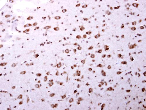
ARA9 antibody detects ARA9 protein at cytosol on mouse middle brain by immunohistochemical analysis. Sample: Paraffin-embedded mouse middle brain. ARA9 antibody dilution: 1:500.Application
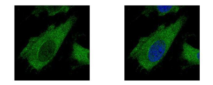
ARA9 antibody detects AIP protein at cytoplasm by confocal immunofluorescent analysis. Sample: HeLa cells were fixed in 4% paraformaldehyde at RT for 15 min. Green: AIP protein stained by ARA9 antibody diluted at 1:500. Blue: Hoechst 33343 staining. [Images captured by Olympus FV10i Confocal Laser Scanning Microscope]Application
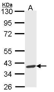
Sample (30 ug of whole cell lysate) A: Molt-4 10% SDS PAGE diluted at 1:1000| Product Name | ARA9 antibody |
|---|---|
| Antibody Type | Primary Antibodies |
| Product description | Rabbit Polyclonal antibody to ARA9 (aryl hydrocarbon receptor interacting protein)The protein encoded by this gene is a receptor for aryl hydrocarbons and a ligand-activated transcription factor. The encoded protein is found in the cytoplasm as part of a multiprotein complex, but upon binding of ligand is transported to the nucleus. This protein can regulate the expression of many xenobiotic metabolizing enzymes. Also, the encoded protein can bind specifically to and inhibit the activity of hepatitis B virus. [provided by RefSeq] |
| Immunogen | Recombinant protein encompassing a sequence within the center region of human ARA9. The exact sequence is proprietary. |
| Clonality | Polyclonal |
|---|---|
| Isotype | IgG |
| Host Species | Rabbit |
| Tested Applications | ICC/IFIHC-PWB |
| *Optimal dilutions/concentrations should be determined by the researcher. | |
| Species Reactivity | HumanMouse |
| Predicted Reactivity | Rat, Chicken, Rhesus Monkey, BovineRat (93%), Chicken (82%), Rhesus Monkey (97%), Bovine (92%) |
| Concentration | 1mg/ml |
| Purification | Affinity purified |
| Gene Synonyms | ARA9 antibody, FKBP16 antibody, FKBP37 antibody, SMTPHN antibody, XAP2 antibody, AIP antibody, aryl hydrocarbon receptor-interacting protein antibody, aryl-hydrocarbon receptor-interacting protein antibody, immunophilin homolog ARA9 antibody, HBV X-associa |
|---|---|
| Molecular Weight(MW) | 38 kDa (note) (3) |
| Cellular Localization | Cytoplasm |
Application
ARA9 antibody detects ARA9 protein at cytosol on mouse middle brain by immunohistochemical analysis. Sample: Paraffin-embedded mouse middle brain. ARA9 antibody dilution: 1:500.
Application
ARA9 antibody detects AIP protein at cytoplasm by confocal immunofluorescent analysis. Sample: HeLa cells were fixed in 4% paraformaldehyde at RT for 15 min. Green: AIP protein stained by ARA9 antibody diluted at 1:500. Blue: Hoechst 33343 staining. [Images captured by Olympus FV10i Confocal Laser Scanning Microscope]
Application
Sample (30 ug of whole cell lysate) A: Molt-4 10% SDS PAGE diluted at 1:1000| Positive Control | Molt-4 |
|---|---|
| Application Notes | *Optimal dilutions/concentrations should be determined by the researcher. |
| Form | Liquid |
|---|---|
| Storage Instructions | Keep as concentrated solution. Aliquot and store at -20ºC or below. Avoid multiple freeze-thaw cycles. |
| Storage Buffer | 0.1M Tris, 0.1M Glycine, 20% Glycerol (pH7). 0.01% Thimerosal was added as a preservative. |


