Application
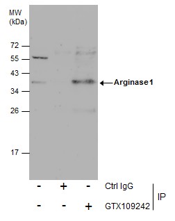
Immunoprecipitation of Arginase 1 protein from HepG2 whole cell extracts using 5 μg of Arginase 1 antibody . Western blot analysis was performed using Arginase 1 antibody . EasyBlot anti-Rabbit IgG was used as a secondary reagent.Application
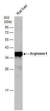
Rat tissue extract (50 µg) was separated by 12% SDS-PAGE, and the membrane was blotted with Arginase 1 antibody diluted at 1:10000. The HRP-conjugated anti-rabbit IgG antibody was used to detect the primary antibody.Application
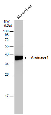
Mouse tissue extract (50 µg) was separated by 12% SDS-PAGE, and the membrane was blotted with Arginase 1 antibody diluted at 1:10000. The HRP-conjugated anti-rabbit IgG antibody was used to detect the primary antibody.Application
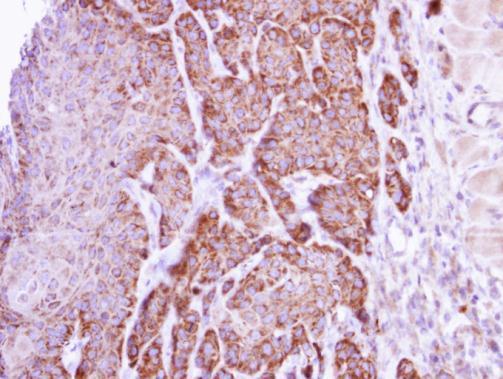
Immunohistochemical analysis of paraffin-embedded Cal27 xenograft, using arginase I antibody at 1:500 dilution.Application
Arginase 1 antibody detects ARG1 protein by flow cytometry analysis. Sample: HepG2 cell. Black: Unlabelled sample was used as a control. Red: Arginase 1 antibody dilution: 1:50. Acquisition of 20,000 events were collected using a Dylight 488-conjugated secondary antibody for FACS analysis.Application
Immunofluorescence analysis of paraformaldehyde-fixed HeLa, using arginase I antibody at 1:200 dilution.Application
Arginase 1 antibody detects Arginase 1 protein by western blot analysis. Various whole cell extracts (30 µg) were separated by 12% SDS-PAGE, and the membrane was blotted with Arginase 1 antibody diluted at 1:1000. The HRP-conjugated anti-rabbit IgG antibody was used to detect the primary antibody.Application
Non-transfected (–) and transfected (+) HepG2 whole cell extracts (30 µg) were separated by 10% SDS-PAGE, and the membrane was blotted with Arginase 1 antibody diluted at 1:1000. The HRP-conjugated anti-rabbit IgG antibody was used to detect the primary antibody.| Product Name | Arginase 1 antibody |
|---|---|
| Antibody Type | Primary Antibodies |
| Product description | Rabbit Polyclonal antibody to Arginase 1 (arginase, liver)Arginase catalyzes the hydrolysis of arginine to ornithine and urea. At least two isoforms of mammalian arginase exist (types I and II) which differ in their tissue distribution, subcellular localization, immunologic crossreactivity and physiologic function. The type I isoform encoded by this gene, is a cytosolic enzyme and expressed predominantly in the liver as a component of the urea cycle. Inherited deficiency of this enzyme results in argininemia, an autosomal recessive disorder characterized by hyperammonemia. [provided by RefSeq] |
| Immunogen | Full length human Arginase 1 Recombinant protein. |
| Clonality | Polyclonal |
|---|---|
| Isotype | IgG |
| Host Species | Rabbit |
| Tested Applications | FACSICC/IFIHC-FIHC-PIPWB |
| *Optimal dilutions/concentrations should be determined by the researcher. | |
| Species Reactivity | HumanMouseRat |
| Predicted Reactivity | Pig, Rabbit, BovinePig (89%), Rabbit (91%), Bovine (90%) |
| Concentration | 1mg/ml |
| Purification | Affinity purified |
| Gene Synonyms | ARG1 antibody , arginase 1 antibody , type I arginase antibody , liver-type arginase antibody , arginase, liver antibody |
|---|---|
| Molecular Weight(MW) | 35 kDa (note) |
| Cellular Localization | Cytoplasm |
Application
Immunoprecipitation of Arginase 1 protein from HepG2 whole cell extracts using 5 μg of Arginase 1 antibody . Western blot analysis was performed using Arginase 1 antibody . EasyBlot anti-Rabbit IgG was used as a secondary reagent.
Application
Rat tissue extract (50 µg) was separated by 12% SDS-PAGE, and the membrane was blotted with Arginase 1 antibody diluted at 1:10000. The HRP-conjugated anti-rabbit IgG antibody was used to detect the primary antibody.
Application
Mouse tissue extract (50 µg) was separated by 12% SDS-PAGE, and the membrane was blotted with Arginase 1 antibody diluted at 1:10000. The HRP-conjugated anti-rabbit IgG antibody was used to detect the primary antibody.
Application
Immunohistochemical analysis of paraffin-embedded Cal27 xenograft, using arginase I antibody at 1:500 dilution.
Application
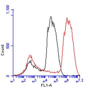
Arginase 1 antibody detects ARG1 protein by flow cytometry analysis. Sample: HepG2 cell. Black: Unlabelled sample was used as a control. Red: Arginase 1 antibody dilution: 1:50. Acquisition of 20,000 events were collected using a Dylight 488-conjugated secondary antibody for FACS analysis.
Application
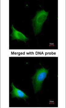
Immunofluorescence analysis of paraformaldehyde-fixed HeLa, using arginase I antibody at 1:200 dilution.
Application
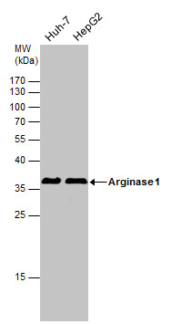
Arginase 1 antibody detects Arginase 1 protein by western blot analysis. Various whole cell extracts (30 µg) were separated by 12% SDS-PAGE, and the membrane was blotted with Arginase 1 antibody diluted at 1:1000. The HRP-conjugated anti-rabbit IgG antibody was used to detect the primary antibody.
Application
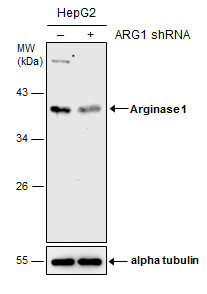
Non-transfected (–) and transfected (+) HepG2 whole cell extracts (30 µg) were separated by 10% SDS-PAGE, and the membrane was blotted with Arginase 1 antibody diluted at 1:1000. The HRP-conjugated anti-rabbit IgG antibody was used to detect the primary antibody.| Positive Control | Huh-7 , HepG2 , mouse liver , rat liver |
|---|---|
| Application Notes | *Optimal dilutions/concentrations should be determined by the researcher. |
| Form | Liquid |
|---|---|
| Storage Instructions | Keep as concentrated solution. Aliquot and store at -20ºC or below. Avoid multiple freeze-thaw cycles. |
| Storage Buffer | 1XPBS, 20% Glycerol (pH7). 0.025% ProClin 300 was added as a preservative. |


