Application
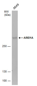
ARID1A antibody detects ARID1A protein by western blot analysis. Whole cell extracts (30 μg) was separated by 5% SDS-PAGE, and the membrane was blotted with ARID1A antibody diluted by 1:1000.Application
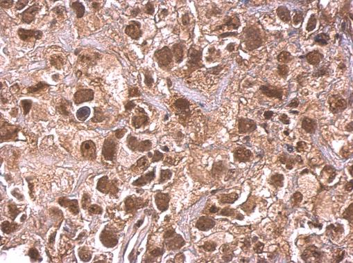
ARID1A antibody detects ARID1A protein at nucleus on human cervical carcinoma by immunohistochemical analysis. Sample: Paraffin-embedded human cervical carcinoma. ARID1A antibody dilution: 1:500.Application
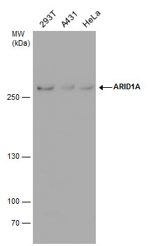
ARID1A antibody detects ARID1A protein by western blot analysis. Various whole cell extracts (30 μg) were separated by 5% SDS-PAGE, and the membrane was blotted with ARID1A antibody diluted by 1:1000.Application
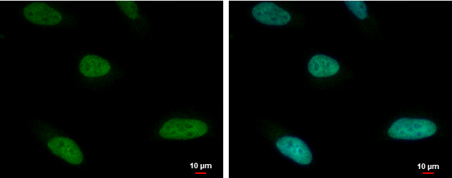
ARID1A antibody detects ARID1A protein at nucleus by immunofluorescent analysis. Sample: HeLa cells were fixed in 4% paraformaldehyde at RT for 15 min. Green: ARID1A protein stained by ARID1A antibody diluted at 1:500. Blue: Hoechst 33342 staining.Application
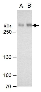
ARID1A antibody detects ARID1A protein by western blot analysis. A. 30 μg HepG2 whole cell extract B. 30 μg HepG2 nuclear extract 5 % SDS-PAGE ARID1A antibody dilution: 1:1000Application
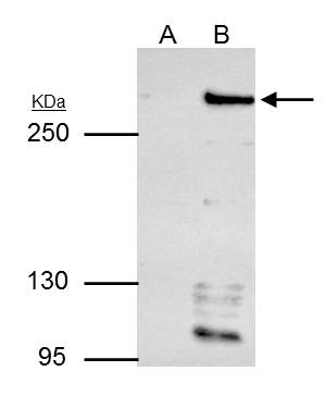
ARID1A antibody immunoprecipitates ARID1A protein in IP experiments. IP samples: HepG2 whole cell extract A. Control with 4 μg of preimmune Rabbit IgG B. Immunoprecipitation of ARID1A protein by 4 μg ARID1A antibody 5 % SDS-PAGE The immunoprecipitated ARID1A protein was detected by ARID1A antibody diluted at 1:500. [EasyBlot anti-rabbit IgG was used as a secondary reagent]Application
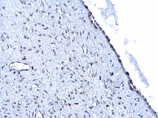
ARID1A antibody detects ARID1A protein at nucleus on human endometrial carcinoma by immunohistochemical analysis. Sample: Paraffin-embedded human endometrial carcinoma. ARID1A antibody diluted at 1:500.| Product Name | ARID1A antibody |
|---|---|
| Antibody Type | Primary Antibodies |
| Product description | Rabbit Polyclonal antibody to ARID1A (AT rich interactive domain 1A (SWI-like))This gene encodes a member of the SWI/SNF family, whose members have helicase and ATPase activities and are thought to regulate transcription of certain genes by altering the chromatin structure around those genes. The encoded protein is part of the large ATP-dependent chromatin remodeling complex SNF/SWI, which is required for transcriptional activation of genes normally repressed by chromatin. It possesses at least two conserved domains that could be important for its function. First, it has a DNA-binding domain that can specifically bind an AT-rich DNA sequence known to be recognized by a SNF/SWI complex at the beta-globin locus. Second, the C-terminus of the protein can stimulate glucocorticoid receptor-dependent transcriptional activation. It is thought that the protein encoded by this gene confers specificity to the SNF/SWI complex and may recruit the complex to its targets through either protein-DNA or protein-protein interactions. Two transcript variants encoding different isoforms have been found for this gene. [provided by RefSeq] |
| Immunogen | Recombinant protein encompassing a sequence within the C-terminus region of human ARID1A. The exact sequence is proprietary. |
| Clonality | Polyclonal |
|---|---|
| Isotype | IgG |
| Host Species | Rabbit |
| Tested Applications | ICC/IFIHC-PIPWB |
| *Optimal dilutions/concentrations should be determined by the researcher. | |
| Species Reactivity | Human |
| Predicted Reactivity | Mouse, Rat, BovineMouse (81%), Rat (81%), Bovine (84%) |
| Concentration | 1mg/ml |
| Purification | Affinity purified |
| Gene Synonyms | BAF250, C1orf4, AT rich interactive domain 1A (SWI-like), B120, P270, SMARCF1, ELD, OSA1, hELD, hOSA1, BAF250a, MRD14, BM029 |
|---|---|
| Target | ARID1A |
| Cellular Localization | Nucleus |
Application
ARID1A antibody detects ARID1A protein by western blot analysis. Whole cell extracts (30 μg) was separated by 5% SDS-PAGE, and the membrane was blotted with ARID1A antibody diluted by 1:1000.
Application
ARID1A antibody detects ARID1A protein at nucleus on human cervical carcinoma by immunohistochemical analysis. Sample: Paraffin-embedded human cervical carcinoma. ARID1A antibody dilution: 1:500.
Application
ARID1A antibody detects ARID1A protein by western blot analysis. Various whole cell extracts (30 μg) were separated by 5% SDS-PAGE, and the membrane was blotted with ARID1A antibody diluted by 1:1000.
Application
ARID1A antibody detects ARID1A protein at nucleus by immunofluorescent analysis. Sample: HeLa cells were fixed in 4% paraformaldehyde at RT for 15 min. Green: ARID1A protein stained by ARID1A antibody diluted at 1:500. Blue: Hoechst 33342 staining.
Application
ARID1A antibody detects ARID1A protein by western blot analysis. A. 30 μg HepG2 whole cell extract B. 30 μg HepG2 nuclear extract 5 % SDS-PAGE ARID1A antibody dilution: 1:1000
Application
ARID1A antibody immunoprecipitates ARID1A protein in IP experiments. IP samples: HepG2 whole cell extract A. Control with 4 μg of preimmune Rabbit IgG B. Immunoprecipitation of ARID1A protein by 4 μg ARID1A antibody 5 % SDS-PAGE The immunoprecipitated ARID1A protein was detected by ARID1A antibody diluted at 1:500. [EasyBlot anti-rabbit IgG was used as a secondary reagent]
Application
ARID1A antibody detects ARID1A protein at nucleus on human endometrial carcinoma by immunohistochemical analysis. Sample: Paraffin-embedded human endometrial carcinoma. ARID1A antibody diluted at 1:500.| Application Notes | *Optimal dilutions/concentrations should be determined by the researcher. |
|---|
| Form | Liquid |
|---|---|
| Storage Instructions | Keep as concentrated solution. Aliquot and store at -20ºC or below. Avoid multiple freeze-thaw cycles. |
| Storage Buffer | 0.2M Tris, 0.2M Glycine, 1mM EGTA, 50% Glycerol (pH7). 0.025% ProClin 300 was added as a preservative. |


