Application
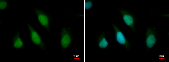
ARID2 antibody detects ARID2 protein at nucleus by immunofluorescent analysis. Sample: HeLa cells were fixed in 4% paraformaldehyde at RT for 15 min. Green: ARID2 protein stained by ARID2 antibody diluted at 1:500. Blue: Hoechst 33342 staining.Application
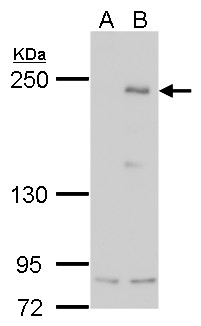
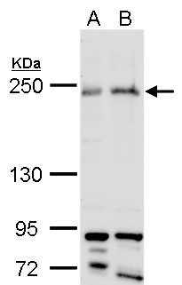
ARID2 antibody detects ARID2 protein by western blot analysis. A. 30 µg 293T B. 30 µg of human ARID2-transfected 293T cells 5% SDS-PAGE ARID2 antibody dilution: 1:5000 The HRP-conjugated anti-rabbit IgG antibody was used to detect the primary antibody.Application
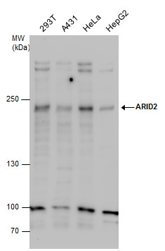
ARID2 antibody detects ARID2 protein by western blot analysis. Various whole cell extracts (30 µg) were separated by 5% SDS-PAGE, and the membrane was blotted with ARID2 antibody diluted by 1:500. The HRP-conjugated anti-rabbit IgG antibody was used to detect the primary antibody.Application
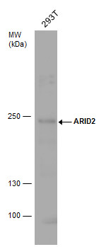
ARID2 antibody detects ARID2 protein by western blot analysis. Whole cell extracts (30 µg) was separated by 5% SDS-PAGE, and the membrane was blotted with ARID2 antibody diluted by 1:500. The HRP-conjugated anti-rabbit IgG antibody was used to detect the primary antibody.Application
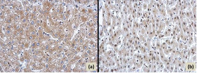
ARID2 antibody detects ARID2 protein on human hepatoma and human normal liver by immunohistochemical analysis. Sample: (a) Paraffin-embedded human hepatoma (b) Paraffin-embedded human normal liver. ARID2 antibody dilution: 1:1000.Application
ARID2 antibody detects ARID2 protein by western blot analysis. A. 30 µg HeLa whole cell lysate/extract B. 30 µg HeLa nuclear lysate/extract 5% SDS-PAGE ARID2 antibody dilution: 1:500 The HRP-conjugated anti-rabbit IgG antibody was used to detect the primary antibody.Application
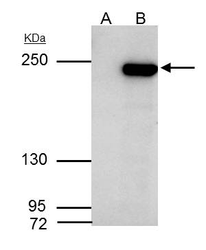
ARID2 antibody immunoprecipitates ARID2 protein in IP experiments. IP samples: HepG2 whole cell extract A. Control with 4 μg of preimmune Rabbit IgG B. Immunoprecipitation of ARID2 protein by 4 μg ARID2 antibody 5 % SDS-PAGE The immunoprecipitated ARID2 protein was detected by ARID2 antibody diluted at 1:500. [EasyBlot anti-rabbit IgG was used as a secondary reagent]| Product Name | ARID2 antibody |
|---|---|
| Antibody Type | Primary Antibodies |
| Product description | Rabbit Polyclonal antibody to ARID2 (AT rich interactive domain 2 (ARID, RFX-like))ARID2 is a subunit of the PBAF chromatin-remodeling complex (see BAF180; MIM 606083), which facilitates ligand-dependent transcriptional activation by nuclear receptors (Yan et al., 2005 [PubMed 15985610]).[supplied by OMIM] |
| Immunogen | Recombinant protein encompassing a sequence within the C-terminus region of human ARID2. The exact sequence is proprietary. |
| Clonality | Polyclonal |
|---|---|
| Isotype | IgG |
| Host Species | Rabbit |
| Tested Applications | ICC/IFIHC-PIPWB |
| *Optimal dilutions/concentrations should be determined by the researcher. | |
| Species Reactivity | Human |
| Predicted Reactivity | MouseMouse (95%) |
| Concentration | 1mg/ml (Please refer to the vial label for the specific concentration) |
| Purification | Affinity purified |
| Gene Synonyms | AT rich interactive domain 2 (ARID, RFX-like), BAF200, p200 |
|---|---|
| Target | ARID2 |
| Cellular Localization | Nucleus |
Application
ARID2 antibody detects ARID2 protein at nucleus by immunofluorescent analysis. Sample: HeLa cells were fixed in 4% paraformaldehyde at RT for 15 min. Green: ARID2 protein stained by ARID2 antibody diluted at 1:500. Blue: Hoechst 33342 staining.
Application
ARID2 antibody detects ARID2 protein by western blot analysis. A. 30 µg 293T B. 30 µg of human ARID2-transfected 293T cells 5% SDS-PAGE ARID2 antibody dilution: 1:5000 The HRP-conjugated anti-rabbit IgG antibody was used to detect the primary antibody.
Application
ARID2 antibody detects ARID2 protein by western blot analysis. Various whole cell extracts (30 µg) were separated by 5% SDS-PAGE, and the membrane was blotted with ARID2 antibody diluted by 1:500. The HRP-conjugated anti-rabbit IgG antibody was used to detect the primary antibody.
Application
ARID2 antibody detects ARID2 protein by western blot analysis. Whole cell extracts (30 µg) was separated by 5% SDS-PAGE, and the membrane was blotted with ARID2 antibody diluted by 1:500. The HRP-conjugated anti-rabbit IgG antibody was used to detect the primary antibody.
Application
ARID2 antibody detects ARID2 protein on human hepatoma and human normal liver by immunohistochemical analysis. Sample: (a) Paraffin-embedded human hepatoma (b) Paraffin-embedded human normal liver. ARID2 antibody dilution: 1:1000.
Application
ARID2 antibody detects ARID2 protein by western blot analysis. A. 30 µg HeLa whole cell lysate/extract B. 30 µg HeLa nuclear lysate/extract 5% SDS-PAGE ARID2 antibody dilution: 1:500 The HRP-conjugated anti-rabbit IgG antibody was used to detect the primary antibody.
Application
ARID2 antibody immunoprecipitates ARID2 protein in IP experiments. IP samples: HepG2 whole cell extract A. Control with 4 μg of preimmune Rabbit IgG B. Immunoprecipitation of ARID2 protein by 4 μg ARID2 antibody 5 % SDS-PAGE The immunoprecipitated ARID2 protein was detected by ARID2 antibody diluted at 1:500. [EasyBlot anti-rabbit IgG was used as a secondary reagent]| Application Notes | *Optimal dilutions/concentrations should be determined by the researcher. |
|---|
| Form | Liquid |
|---|---|
| Storage Instructions | Keep as concentrated solution. Aliquot and store at -20ºC or below. Avoid multiple freeze-thaw cycles. |
| Storage Buffer | 1XPBS, 20% Glycerol (pH7). 0.01% Thimerosal was added as a preservative. |


