Application
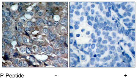
Immunohistochemical analysis of paraffin-embedded human breast carcinoma tissue using ASK1 (phospho-Ser83) antibodyApplication
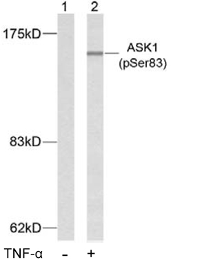
Western blot analysis of extracts from K562 cells using ASK1 (phospho-Ser83) antibodyApplication
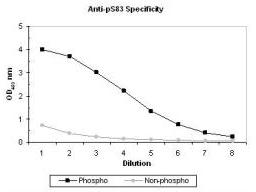
ELISA results of purified polyclonal anti-pS83 ASK-1 (aa 76-87) antibody tested against BSA conjugates of non-phospho and phospho forms of immunizing peptide. Each well was coated with 0.1 mg of conjugate. The starting dilution of antibody was 1:1,000 and each point on the X-axis represents a 2-fold dilution. HRP conjugated Gt-a-Rabbit IgG H&L and TMB substrate were used for detection.Application
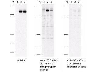
Western blot of anti-pS83 ASK1 antibodies shows specificity for phosphorylated human ASK1. Anti-pS83 (aa 76-87) antibody was tested by western blot against Cos-7 cell lysates after transient transfection with 1) vector only, 2) recombinant HA-ASK1, and 3) recombinant human HA-ASK1 where S83 was substituted with an alanine residue. Cells were lysed 24 h post-transfection in 200 ?L of 1x SDS-sample buffer, heated at 96?C for 5', and vortexed for 30 sec. Samples (10 uL each) were separated on a 12% SDS-PAGE gel and transferred to PVDF (Millipore) followed by blocking for 45' using TTBS supplemented with 5% non-fat dry milk. All incubations were performed at room temperature. In panel a) all samples were incubated with anti-HA antibody. This blot demonstrates both recombinant transfections express rASK1. In panel b) all samples were incubated with anti-pS83 ASK1. Lane 2 shows strong specific staining of ASK1. Lane 3, where S83 was replaced with alanine, shows greatly diminished staining. In panel c) all samples were incubated with anti-pS83 ASK1 antibody as before except the antibody was pre-incubated with phospho peptide prior to membrane incubation. No staining is observed after phospho peptide blocking occurs.| Product Name | ASK1 (phospho Ser83) antibody |
|---|---|
| Antibody Type | Primary Antibodies |
| Product description | Rabbit polyclonal to ASK1 ( phospho S83 )Mitogen-activated protein (MAP) kinase cascades are activated in response to various extracellular stimuli including cytokines, growth factors and environmental stresses. A novel MAP kinase kinase kinase (MAPKKK) was recently identified and designated ASK1 (for apoptosis signal-regulating kinase 1) and MAPKKK5. ASK1 activates two different subgroups of MAPKK, MKK4 and MKK6, which in turn activate c-Jun N-terminal kinase (JNK) and p38 MAP kinase, respectively. ASK1/MAPKKK5 is activated by TNFR and Fas through the interaction with members of the TRAF family and Fas-associated protein Daxx. Overexpression of ASK1 induces apoptotic cell death, and a catalytically inactive form of ASK1 inhibits TNF-a induced apoptosis. ASK1 is expressed in variety of human and mouse tissues. |
| Immunogen | The antiserum was produced against synthesized phosphopeptide derived from human ASK1 around the phosphorylation site of serine 83 (G-S-S^P-V-G). |
| Modification | p-Ser83 |
| Clonality | Polyclonal |
|---|---|
| Isotype | IgG |
| Host Species | Rabbit |
| Tested Applications | ELISAIHC-PWB |
| For WB: Use at a dilution of 1:500-1:1000. Predicted molecular weight: 155 kDa. For IHC-P: Use at a dilution of 1:50-1:1000. Optimal dilutions/concentrations should be determined by the end user.: |
|
| Species Reactivity | Human |
| Concentration | 1mg/ml (Please refer to the vial label for the specific concentration) |
| Purification | Affinity purified |
| Gene Synonyms | ASK1 (phospho S83) ASK1 (phospho Ser83) ASK1 (pS83) ASK1 (pSer83) ASK1 phospho S83 ASK1 phospho Ser83 Phospho ASK1 (S83) Phospho ASK1 (Ser83) Phospho ASK1 (pS83) Phospho ASK1 (pSer83) |
|---|---|
| Target | ASK1 |
| Tissue Specificity | ASK1 (phospho-Ser83) antibody detects endogenous levels of ASK1 only when phosphorylated at serine 83. |
| Cellular Localization | Cytoplasmic |
Application
Immunohistochemical analysis of paraffin-embedded human breast carcinoma tissue using ASK1 (phospho-Ser83) antibody
Application
Western blot analysis of extracts from K562 cells using ASK1 (phospho-Ser83) antibody
Application
ELISA results of purified polyclonal anti-pS83 ASK-1 (aa 76-87) antibody tested against BSA conjugates of non-phospho and phospho forms of immunizing peptide. Each well was coated with 0.1 mg of conjugate. The starting dilution of antibody was 1:1,000 and each point on the X-axis represents a 2-fold dilution. HRP conjugated Gt-a-Rabbit IgG H&L and TMB substrate were used for detection.
Application
Western blot of anti-pS83 ASK1 antibodies shows specificity for phosphorylated human ASK1. Anti-pS83 (aa 76-87) antibody was tested by western blot against Cos-7 cell lysates after transient transfection with 1) vector only, 2) recombinant HA-ASK1, and 3) recombinant human HA-ASK1 where S83 was substituted with an alanine residue. Cells were lysed 24 h post-transfection in 200 ?L of 1x SDS-sample buffer, heated at 96?C for 5', and vortexed for 30 sec. Samples (10 uL each) were separated on a 12% SDS-PAGE gel and transferred to PVDF (Millipore) followed by blocking for 45' using TTBS supplemented with 5% non-fat dry milk. All incubations were performed at room temperature. In panel a) all samples were incubated with anti-HA antibody. This blot demonstrates both recombinant transfections express rASK1. In panel b) all samples were incubated with anti-pS83 ASK1. Lane 2 shows strong specific staining of ASK1. Lane 3, where S83 was replaced with alanine, shows greatly diminished staining. In panel c) all samples were incubated with anti-pS83 ASK1 antibody as before except the antibody was pre-incubated with phospho peptide prior to membrane incubation. No staining is observed after phospho peptide blocking occurs.| Application Notes | For WB: Use at a dilution of 1:500-1:1000. Predicted molecular weight: 155 kDa. For IHC-P: Use at a dilution of 1:50-1:1000. Optimal dilutions/concentrations should be determined by the end user.: |
|---|
| Form | Liquid |
|---|---|
| Storage Instructions | Upon Receipt - Keep as concentrated solution. Aliquot and store at -20ºC or below. Avoid freeze-thaw cycles. |
| Storage Buffer | Phosphate buffered saline, pH 7.3, 50% glycerol and 150mM NaCl containing 0.02% sodium azide |


