
Application
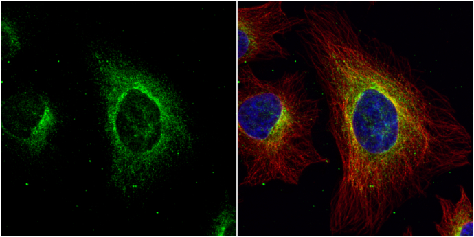
Immunofluorescence analysis of paraformaldehyde-fixed HeLa, using Argininosuccinate Lyase antibody at 1:200 dilution.Application
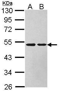
Sample (30 µg of whole cell lysate) A: HeLa B: HepG2 10% SDS PAGE diluted at 1:1000 The HRP-conjugated anti-rabbit IgG antibody was used to detect the primary antibody.Application
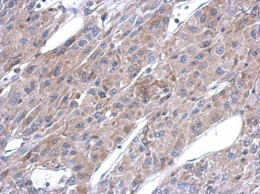
Immunohistochemical analysis of paraffin-embedded U87 xenograft, using ASL antibody at 1:500 dilution.Application
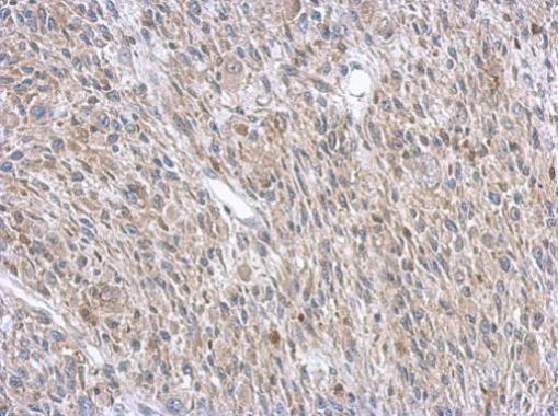
Immunohistochemical analysis of paraffin-embedded C2C12 xenograft, using ASL antibody at 1:500 dilution.Application
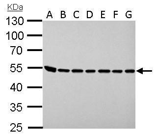
ASL antibody detects ASL protein by western blot analysis. A. 30 µg Neuro2A whole lysate/extract B. 30 µg GL261 whole cell lysate/extract C. 30 µg C8D30 whole cell lysate/extract D. 30 µg NIH-3T3 whole cell lysate/extract E. 30 µg BCL-1 whole cell lysate/extract F. 30 µg Raw264.7 whole cell lysate/extract G. 30 µg C2C12 whole cell lysate/extract 10% SDS-PAGE ASL antibody dilution: 1:1000 The HRP-conjugated anti-rabbit IgG antibody was used to detect the primary antibody.Application
ASL antibody detects ASL protein at cytoplasm by immunofluorescent analysis. Sample: HeLa cells were fixed in 4% paraformaldehyde at RT for 15 min. Green: ASL protein stained by ASL antibody diluted at 1:500. Red: alpha Tubulin, a cytoskeleton marker, stained by alpha Tubulin antibody [GT114] diluted at 1:1000. Blue: Hoechst 33342 staining.| Product Name | ASL antibody |
|---|---|
| Antibody Type | Primary Antibodies |
| Product description | Rabbit Polyclonal antibody to ASL (argininosuccinate lyase)This gene encodes a member of the lyase 1 family. The encoded protein forms a cytosolic homotetramer and primarily catalyzes the reversible hydrolytic cleavage of argininosuccinate into arginine and fumarate, an essential step in the liver in detoxifying ammonia via the urea cycle. Mutations in this gene result in the autosomal recessive disorder argininosuccinic aciduria, or argininosuccinic acid lyase deficiency. A nontranscribed pseudogene is also located on the long arm of chromosome 22. Alternatively spliced transcript variants encoding different isoforms have been described. [provided by RefSeq] |
| Immunogen | Recombinant protein encompassing a sequence within the center region of human ASL. The exact sequence is proprietary. |
| Clonality | Polyclonal |
|---|---|
| Isotype | IgG |
| Host Species | Rabbit |
| Tested Applications | ICC/IFIHC-PWB |
| *Optimal dilutions/concentrations should be determined by the researcher.
: |
|
| Species Reactivity | HumanMouse |
| Predicted Reactivity | Rat, BovineRat (88%), Bovine (95%) |
| Concentration | 1mg/ml (Please refer to the vial label for the specific concentration) |
| Purification | Affinity purified |
| Gene Synonyms | ASAL antibody ASL antibody arginosuccinase antibody argininosuccinase antibody argininosuccinate lyase antibody |
|---|---|
| Molecular Weight(MW) | 52 kDa (note) (5) |
Application
Immunofluorescence analysis of paraformaldehyde-fixed HeLa, using Argininosuccinate Lyase antibody at 1:200 dilution.
Application
Sample (30 µg of whole cell lysate) A: HeLa B: HepG2 10% SDS PAGE diluted at 1:1000 The HRP-conjugated anti-rabbit IgG antibody was used to detect the primary antibody.
Application
Immunohistochemical analysis of paraffin-embedded U87 xenograft, using ASL antibody at 1:500 dilution.
Application
Immunohistochemical analysis of paraffin-embedded C2C12 xenograft, using ASL antibody at 1:500 dilution.
Application
ASL antibody detects ASL protein by western blot analysis. A. 30 µg Neuro2A whole lysate/extract B. 30 µg GL261 whole cell lysate/extract C. 30 µg C8D30 whole cell lysate/extract D. 30 µg NIH-3T3 whole cell lysate/extract E. 30 µg BCL-1 whole cell lysate/extract F. 30 µg Raw264.7 whole cell lysate/extract G. 30 µg C2C12 whole cell lysate/extract 10% SDS-PAGE ASL antibody dilution: 1:1000 The HRP-conjugated anti-rabbit IgG antibody was used to detect the primary antibody.
Application
ASL antibody detects ASL protein at cytoplasm by immunofluorescent analysis. Sample: HeLa cells were fixed in 4% paraformaldehyde at RT for 15 min. Green: ASL protein stained by ASL antibody diluted at 1:500. Red: alpha Tubulin, a cytoskeleton marker, stained by alpha Tubulin antibody [GT114] diluted at 1:1000. Blue: Hoechst 33342 staining.| Positive Control | HeLa , HepG2 , Neuro2A , GL261 , C8D30 , NIH-3T3 , BCL-1 , Raw264.7 , C2C12 |
|---|---|
| Application Notes | *Optimal dilutions/concentrations should be determined by the researcher.
: |
| Form | Liquid |
|---|---|
| Storage Instructions | Keep as concentrated solution. Aliquot and store at -20ºC or below. Avoid multiple freeze-thaw cycles. |
| Storage Buffer | 0.1M Tris, 0.1M Glycine, 20% Glycerol (pH7). 0.01% Thimerosal was added as a preservative. |


