Application
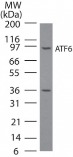
Western blot analysis of ATF6 in NIH3T3 cell lysate using IMG-273 at 3 ug/ml. A band corresponding to full-length ATF6 was detected. We have not characterized the ~36 kDa observed band; it may be an ATF6 breakdown/cleavage product.Application
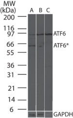
Western blot analysis of ATF6 in mouse liver tissue enriched using 10088K using 3 ug/ml of IMG-273 (anti-ATF6) and 0.25 ug/ml (anti-GAPDH loading control). Lane A contains 20 ugs of whole mouse liver lysate, lane B contains 20 ugs of total ER fraction, and lane C contains 20 ugs of rough ER fraction. The ATF6* band may represent under glycosylated or cleaved/active ATF6.Application
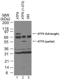
Western blot analysis of ATF6. Lane 1, 293 cells transfected with full-length ATF6.* Lane 2, 293 cells transfected with partial length ATF6 (amino acids 1-373).* Lane 3, Untransfected 293 cells. Western blots were probed with 4 ug/ml of the ATF6 monoclonal antibody, followed by an HRP-conjugated second step (20101) and visualized with PicoTect Western Blot Chemiluminescence Substrate (10087K). Film was exposed for 1 min. The top arrow corresponds to the ~90 kDa form of ATF6 described as full-length in the literature. *The human full-length and partial length ATF6 plasmids are described in Luo and Lee (2002).| Product Name | ATF6 antibody |
|---|---|
| Antibody Type | Primary Antibodies |
| Product description | Mouse monoclonal [70B1413] to ATF6ATF6 is a transcription factor that acts during endoplasmic reticulum stress by activating unfolded protein response target genes. It binds DNA on the 5'-CCAC[GA]-3'half of the ER stress response element (ERSE) (5'-CCAAT-N(9)-CCAC[GA]-3') and of ERSE II (5'-ATTGG-N-CCACG-3'). Binding to ERSE requires binding of NF-Y to ERSE. ATF6 could also be involved in activation of transcription by the serum response factor. ATF6 exists as a homodimer and heterodimer with ATF6 beta. The dimer interacts with the nuclear transcription factor Y (NF-Y) trimer through direct binding to NF-Y subunit C (NF-YC). It Interacts also with the transcription factors GTF2I, YY1 and SRF. Under ER stress the cleaved N-terminal cytoplasmic domain translocates into the nucleus. The basic domain of ATF6 functions as a nuclear localization signal and the basic leucine-zipper domain is sufficient for association with the NF-Y trimer and binding to ERSE. During the unfolded protein response an approximately 50 kDa fragment containing the cytoplasmic transcription factor domain is released by proteolysis. The cleavage seems to be performed sequentially by site-1 and site-2 proteases. ATF6 is N-glycosylated, phosphorylated in vitro by MAPK14/P38MAPK and belongs to the bZIP family. |
| Immunogen | Full length protein (Human). |
| Clonality | Monoclonal |
|---|---|
| Isotype | IgG1 |
| Clone Number | 70B1413 |
| Host Species | Mouse |
| Tested Applications | WB |
| WB: Use at a concentration of 0.5 - 2.0 μg/ml. Detects a band of approximately 90 kDa. Optimal dilutions/concentrations should be determined by the end user.: |
|
| Species Reactivity | Human |
| Concentration | 0.5mg/ml |
| Purification | Affinity purified |
| Gene Synonyms | 605537 ATF6 22926 P18850 |
|---|---|
| Target | ATF6 |
| Tissue Specificity | This antibody only recognizes the 90kDa form of ATF6. |
Application
Western blot analysis of ATF6 in NIH3T3 cell lysate using IMG-273 at 3 ug/ml. A band corresponding to full-length ATF6 was detected. We have not characterized the ~36 kDa observed band; it may be an ATF6 breakdown/cleavage product.
Application
Western blot analysis of ATF6 in mouse liver tissue enriched using 10088K using 3 ug/ml of IMG-273 (anti-ATF6) and 0.25 ug/ml (anti-GAPDH loading control). Lane A contains 20 ugs of whole mouse liver lysate, lane B contains 20 ugs of total ER fraction, and lane C contains 20 ugs of rough ER fraction. The ATF6* band may represent under glycosylated or cleaved/active ATF6.
Application
Western blot analysis of ATF6. Lane 1, 293 cells transfected with full-length ATF6.* Lane 2, 293 cells transfected with partial length ATF6 (amino acids 1-373).* Lane 3, Untransfected 293 cells. Western blots were probed with 4 ug/ml of the ATF6 monoclonal antibody, followed by an HRP-conjugated second step (20101) and visualized with PicoTect Western Blot Chemiluminescence Substrate (10087K). Film was exposed for 1 min. The top arrow corresponds to the ~90 kDa form of ATF6 described as full-length in the literature. *The human full-length and partial length ATF6 plasmids are described in Luo and Lee (2002).| Positive Control | 293 whole cell extract |
|---|---|
| Application Notes | WB: Use at a concentration of 0.5 - 2.0 μg/ml. Detects a band of approximately 90 kDa. Optimal dilutions/concentrations should be determined by the end user.: |
| Form | Liquid |
|---|---|
| Storage Instructions | Keep as concentrated solution. Store at 4ºC short term. For extended storage aliquot and store at -20ºC or below. Avoid freeze-thaw cycles. |
| Storage Buffer | Preservative: 0.02% Sodium Azide; Constituents: PBS |


