Application
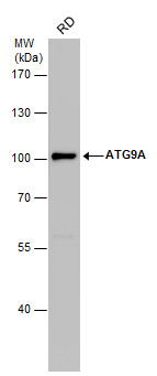
ATG9A antibody detects ATG9A protein by western blot analysis. Whole cell extracts (30 µg) was separated by 7.5% SDS-PAGE, and the membrane was blotted with ATG9A antibody diluted by 1:1000. The HRP-conjugated anti-rabbit IgG antibody was used to detect the primary antibody.Application
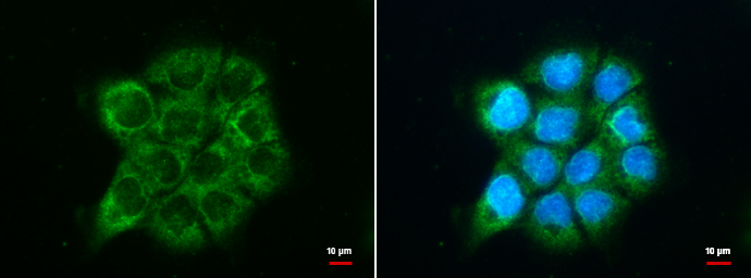
ATG9A antibody detects ATG9A protein at cytoplasm by immunofluorescent analysis. Sample: A431 cells were fixed in 100% MeOH for 5 min. Green: ATG9A protein stained by ATG9A antibody diluted at 1:500. Blue: Hoechst 33342 staining.Application
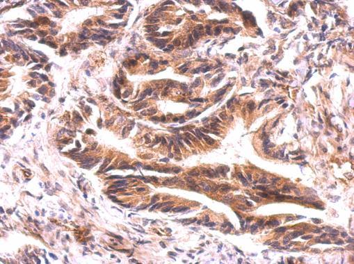
ATG9A antibody detects ATG9A protein on colon carcinoma by immunohistochemical analysis. Sample: Paraffin-embedded colon carcinoma. ATG9A antibody dilution: 1:500.Application
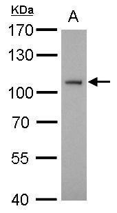
ATG9A antibody detects ATG9A protein by western blot analysis. A. 30 µg BCL-1 whole cell lysate/extract 7.5% SDS-PAGE ATG9A antibody dilution: 1:1000 The HRP-conjugated anti-rabbit IgG antibody was used to detect the primary antibody.Application
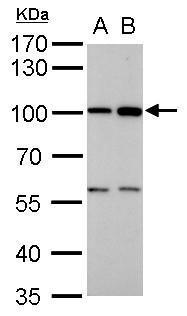
ATG9A antibody detects ATG9A protein by western blot analysis. A. 30 µg PC-12 whole cell lysate/extract B. 30 µg Rat-2 whole cell lysate/extract 7.5% SDS-PAGE ATG9A antibody dilution: 1:1000 The HRP-conjugated anti-rabbit IgG antibody was used to detect the primary antibody.Application
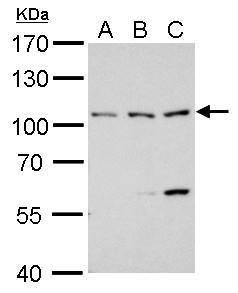
ATG9A antibody detects ATG9A protein by western blot analysis. A. 30 µg 293T whole cell lysate/extract B. 30 µg A431 whole cell lysate/extract C. 30 µg HeLa whole cell lysate/extract 7.5% SDS-PAGE ATG9A antibody dilution: 1:1000 The HRP-conjugated anti-rabbit IgG antibody was used to detect the primary antibody.Application
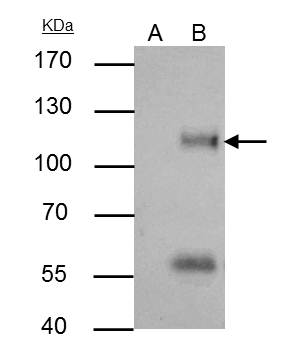
ATG9A antibody immunoprecipitates ATG9A protein in IP experiments. IP samples: HeLa whole cell extract A. Control with 4 μg of preimmune Rabbit IgG B. Immunoprecipitation of ATG9A protein by 4 μg ATG9A antibody 7.5 % SDS-PAGE The immunoprecipitated ATG9A protein was detected by ATG9A antibody diluted at 1:500. [EasyBlot anti-rabbit IgG was used as a secondary reagent]| Product Name | ATG9A antibody |
|---|---|
| Antibody Type | Primary Antibodies |
| Product description | Rabbit Polyclonal antibody to ATG9A (ATG9 autophagy related 9 homolog A (S. cerevisiae))Plays a role in autophagy. |
| Immunogen | Carrier-protein conjugated synthetic peptide encompassing a sequence within the C-terminus region of human ATG9A. The exact sequence is proprietary. |
| Clonality | Polyclonal |
|---|---|
| Isotype | IgG |
| Host Species | Rabbit |
| Tested Applications | ICC/IFIHC-PIPWB |
| *Optimal dilutions/concentrations should be determined by the researcher.
: |
|
| Species Reactivity | HumanMouseRat |
| Predicted Reactivity | Pig, Rhesus Monkey, BovinePig (100%), Rhesus Monkey (100%), Bovine (100%) |
| Concentration | 1mg/ml (Please refer to the vial label for the specific concentration) |
| Purification | Affinity purified |
| Gene Synonyms | APG9L1 antibody MGD3208 antibody mATG9 antibody ATG9A antibody APG9-like 1 antibody autophagy 9-like 1 protein antibody autophagy-related protein 9A antibody APG9 autophagy 9-like 1 antibody ATG9 autophagy related 9 homolog A (S. cerevisiae) antibo |
|---|---|
| Molecular Weight(MW) | 94 kDa (note) (2) |
| Cellular Localization | Cytoplasmic vesicle , autophagosome membrane |
Application
ATG9A antibody detects ATG9A protein by western blot analysis. Whole cell extracts (30 µg) was separated by 7.5% SDS-PAGE, and the membrane was blotted with ATG9A antibody diluted by 1:1000. The HRP-conjugated anti-rabbit IgG antibody was used to detect the primary antibody.
Application
ATG9A antibody detects ATG9A protein at cytoplasm by immunofluorescent analysis. Sample: A431 cells were fixed in 100% MeOH for 5 min. Green: ATG9A protein stained by ATG9A antibody diluted at 1:500. Blue: Hoechst 33342 staining.
Application
ATG9A antibody detects ATG9A protein on colon carcinoma by immunohistochemical analysis. Sample: Paraffin-embedded colon carcinoma. ATG9A antibody dilution: 1:500.
Application
ATG9A antibody detects ATG9A protein by western blot analysis. A. 30 µg BCL-1 whole cell lysate/extract 7.5% SDS-PAGE ATG9A antibody dilution: 1:1000 The HRP-conjugated anti-rabbit IgG antibody was used to detect the primary antibody.
Application
ATG9A antibody detects ATG9A protein by western blot analysis. A. 30 µg PC-12 whole cell lysate/extract B. 30 µg Rat-2 whole cell lysate/extract 7.5% SDS-PAGE ATG9A antibody dilution: 1:1000 The HRP-conjugated anti-rabbit IgG antibody was used to detect the primary antibody.
Application
ATG9A antibody detects ATG9A protein by western blot analysis. A. 30 µg 293T whole cell lysate/extract B. 30 µg A431 whole cell lysate/extract C. 30 µg HeLa whole cell lysate/extract 7.5% SDS-PAGE ATG9A antibody dilution: 1:1000 The HRP-conjugated anti-rabbit IgG antibody was used to detect the primary antibody.
Application
ATG9A antibody immunoprecipitates ATG9A protein in IP experiments. IP samples: HeLa whole cell extract A. Control with 4 μg of preimmune Rabbit IgG B. Immunoprecipitation of ATG9A protein by 4 μg ATG9A antibody 7.5 % SDS-PAGE The immunoprecipitated ATG9A protein was detected by ATG9A antibody diluted at 1:500. [EasyBlot anti-rabbit IgG was used as a secondary reagent]| Positive Control | 293T , A431 , HeLa , RD |
|---|---|
| Application Notes | *Optimal dilutions/concentrations should be determined by the researcher.
: |
| Form | Liquid |
|---|---|
| Storage Instructions | Keep as concentrated solution. Aliquot and store at -20ºC or below. Avoid multiple freeze-thaw cycles. |
| Storage Buffer | 1XPBS, 20% Glycerol (pH7). 0.01% Thimerosal was added as a preservative. |


