Application
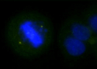
Overnight nocodazole treated Hela cells stained with purified rabbit polyclonal antibody against Thr288 phosphorylated Aurora A, followed by Alexa Fluor 488-conjugated goat anti-rabbit IgG and DAPI.Application
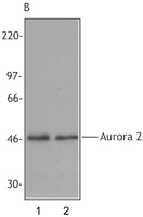
Panel B: The blot shown in Panel A was stripped and re-probed with pan-Aurora specific polyclonal antibody (Poly6033) to verify equal loading of Aurora A in the extracts. Proteins were visualized using a donkey anti-rabbit secondary conjugated to HRP and a chemiluminescence detection system.Application
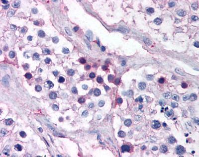
Human Testis (formalin-fixed, paraffin-embedded) stained with AURKA antibody at 1:50 followed by biotinylated goat anti-rabbit IgG secondary antibody LS-D1, alkaline phosphatase-streptavidin and chromogen.Application
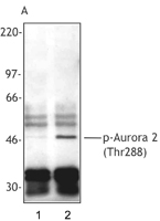
Panel A: Extracts from untreated Hela cells (Lane 1) or overnight nocodazole-treated Hela cells (Lane 2) were immunoprecipitated with a pan-Aurora A monoclonal antibody (clone 35C1), resolved by electrophoresis, transferred to nitrocellulose and probed with rabbit polyclonal antibody against Thr288 phosphorylated Aurora A (Poly6187). Proteins were visualized using a donkey anti-rabbit secondary conjugated to HRP and a chemiluminescence detection system.| Product Name | Aurora A (phospho Thr288) antibody |
|---|---|
| Antibody Type | Primary Antibodies |
| Product description | Rabbit polyclonal antibody to Aurora A (phospho Thr288)The protein encoded by this gene is a cell cycle-regulated kinase that appears to be involved in microtubule formation and/or stabilization at the spindle pole during chromosome segregation. The encoded protein is found at the centrosome in interphase cells and at the spindle poles in mitosis. This gene may play a role in tumor development and progression. A processed pseudogene of this gene has been found on chromosome 1, and an unprocessed pseudogene has been found on chromosome 10. Multiple transcript variants encoding the same protein have been found for this gene. [provided by RefSeq] |
| Immunogen | Synthetic peptide |
| Modification | p-Thr288 |
| Clonality | Polyclonal |
|---|---|
| Isotype | IgG |
| Host Species | Rabbit |
| Tested Applications | ICC/IFIHC-PWB |
| WB, IHC-P (1:50): |
|
| Species Reactivity | Human |
| Concentration | 1mg/ml |
| Purification | Affinity purified |
| Gene Synonyms | Aurora A Aurora related kinase 1 AuroraA Aurorarelated kinase 1 AURKA (phospho T288) AURKA (phospho Thr288) AURKA (pT288) AURKA (pThr288) AURKA phospho T288 AURKA phospho Thr288 Phospho AURKA (T288) Phospho AURKA (Thr288) Phospho AURKA (pT288) |
|---|---|
| Target | AURKA |
| Tissue Specificity | Modified peptide |
Application
Overnight nocodazole treated Hela cells stained with purified rabbit polyclonal antibody against Thr288 phosphorylated Aurora A, followed by Alexa Fluor 488-conjugated goat anti-rabbit IgG and DAPI.
Application
Panel B: The blot shown in Panel A was stripped and re-probed with pan-Aurora specific polyclonal antibody (Poly6033) to verify equal loading of Aurora A in the extracts. Proteins were visualized using a donkey anti-rabbit secondary conjugated to HRP and a chemiluminescence detection system.
Application
Human Testis (formalin-fixed, paraffin-embedded) stained with AURKA antibody at 1:50 followed by biotinylated goat anti-rabbit IgG secondary antibody LS-D1, alkaline phosphatase-streptavidin and chromogen.
Application
Panel A: Extracts from untreated Hela cells (Lane 1) or overnight nocodazole-treated Hela cells (Lane 2) were immunoprecipitated with a pan-Aurora A monoclonal antibody (clone 35C1), resolved by electrophoresis, transferred to nitrocellulose and probed with rabbit polyclonal antibody against Thr288 phosphorylated Aurora A (Poly6187). Proteins were visualized using a donkey anti-rabbit secondary conjugated to HRP and a chemiluminescence detection system.| Application Notes | WB, IHC-P (1:50): |
|---|
| Form | Liquid |
|---|---|
| Storage Instructions | +4°C or -20°C, Avoid repeated freezing and thawing. |
| Storage Buffer | Phosphate-buffered solution, pH 7.2, 0.09% sodium azide, 50% glycerol. |


