Application
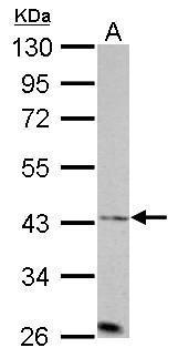
Aurora A antibody [C3], C-term detects AURKA protein by western blot analysis. A. 30 µg PC-12 whole cell lysate/extract 10% SDS-PAGE Aurora A antibody [C3], C-term dilution: 1:1000 The HRP-conjugated anti-rabbit IgG antibody was used to detect the primary antibody.Application
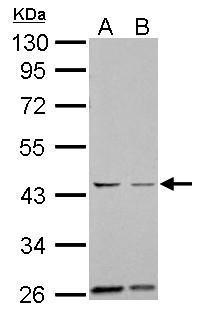
Aurora A antibody [C3], C-term detects AURKA protein by western blot analysis. A. 30 µg Neuro2A whole cell lysate/extract B. 30 µg GL261 whole cell lysate/extract 10% SDS-PAGE Aurora A antibody [C3], C-term dilution: 1:1000 The HRP-conjugated anti-rabbit IgG antibody was used to detect the primary antibody.Application
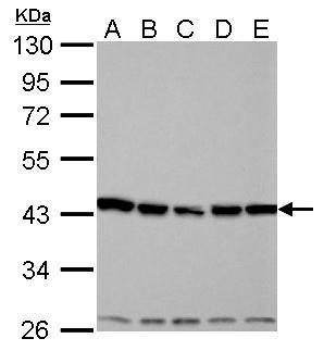
Aurora A antibody [C3], C-term detects AURKA protein by western blot analysis. A. 30 µg 293T whole cell lysate/extract B. 30 µg A431 whole cell lysate/extract C. 30 µg HeLa whole cell lysate/extract D. 30 µg HepG2 whole cell lysate/extract E. 30 µg A375 whole cell lysate/extract 10% SDS-PAGE Aurora A antibody [C3], C-term dilution: 1:1000 The HRP-conjugated anti-rabbit IgG antibody was used to detect the primary antibody.Application
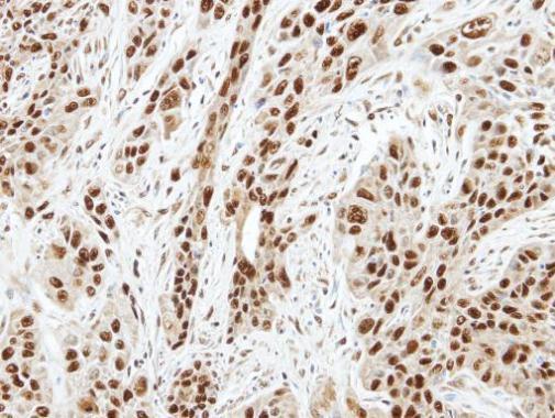
Immunohistochemical analysis of paraffin-embedded TW2.6 xenograft, using Aurora A antibody at 1:100 dilution.Application
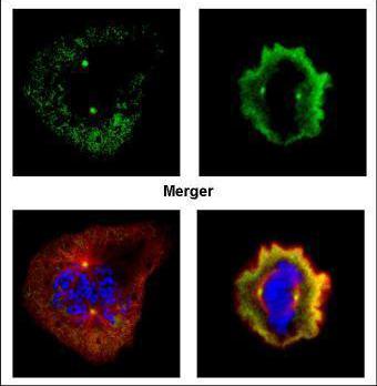
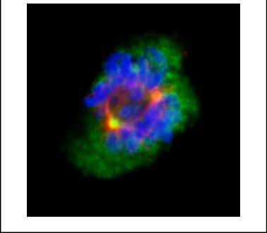
Confocal immunofluorescence analysis (Olympus FV10i) of paraformaldehyde-fixed U2OS, using Aurora A antibody (Green) at 1:500 dilution. Alpha-tubulin filaments were labeled with (Red) at 1:100.Application
Confocal immunofluorescence analysis (Olympus FV10i) of methanol-fixed 293T, using Aurora A antibody (Green) at 1:100 dilution. Alpha-tubulin filaments were labeled with (Red) at 1:2500.Application
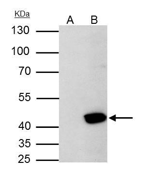
Aurora A antibody [C3], C-term immunoprecipitates Aurora A protein in IP experiments. IP samples: 293T whole cell extract A. Control with 4 μg of preimmune Rabbit IgG B. Immunoprecipitation of Aurora A protein by 4 μg Aurora A antibody [C3], C-term 10 % SDS-PAGE The immunoprecipitated Aurora A protein was detected by Aurora A antibody [C3], C-term diluted at 1:500. [EasyBlot anti-rabbit IgG was used as a secondary reagent]| Product Name | Aurora A antibody , C-term |
|---|---|
| Antibody Type | Primary Antibodies |
| Product description | Rabbit Polyclonal antibody to Aurora A (aurora kinase A)The protein encoded by this gene is a cell cycle-regulated kinase that appears to be involved in microtubule formation and/or stabilization at the spindle pole during chromosome segregation. The encoded protein is found at the centrosome in interphase cells and at the spindle poles in mitosis. This gene may play a role in tumor development and progression. A processed pseudogene of this gene has been found on chromosome 1, and an unprocessed pseudogene has been found on chromosome 10. Multiple transcript variants encoding the same protein have been found for this gene. [provided by RefSeq] |
| Immunogen | Carrier-protein conjugated synthetic peptide encompassing a sequence within the C-terminus region of human Aurora A. The exact sequence is proprietary. |
| Modification | Acetylated |
| Clonality | Polyclonal |
|---|---|
| Isotype | IgG |
| Host Species | Rabbit |
| Tested Applications | ICC/IFIHC-PIPWB |
| WB:1:500~1:2000 IP:1:100~1:500 ICC/IF:1:50~1:100 IHC-p:1:50~1:200 |
|
| Species Reactivity | HumanMouseRat |
| Predicted Reactivity | ChimpanzeeChimpanzee (100%) |
| Concentration | 1mg/ml (Please refer to the vial label for the specific concentration) |
| Purification | Affinity purified |
| Gene Synonyms | AIK antibody ARK1 antibody AURA antibody AURORA2 antibody BTAK antibody MGC34538 antibody STK15 antibody STK6 antibody STK7 antibody AURKA antibody protein phosphatase 1 regulatory subunit 47 antibody ARK-1 antibody aurora/IPL1-like kinase ant |
|---|---|
| Molecular Weight(MW) | 46 kDa (note) (6) |
| Cellular Localization | Centrosome , Spindle |
Application
Aurora A antibody [C3], C-term detects AURKA protein by western blot analysis. A. 30 µg PC-12 whole cell lysate/extract 10% SDS-PAGE Aurora A antibody [C3], C-term dilution: 1:1000 The HRP-conjugated anti-rabbit IgG antibody was used to detect the primary antibody.
Application
Aurora A antibody [C3], C-term detects AURKA protein by western blot analysis. A. 30 µg Neuro2A whole cell lysate/extract B. 30 µg GL261 whole cell lysate/extract 10% SDS-PAGE Aurora A antibody [C3], C-term dilution: 1:1000 The HRP-conjugated anti-rabbit IgG antibody was used to detect the primary antibody.
Application
Aurora A antibody [C3], C-term detects AURKA protein by western blot analysis. A. 30 µg 293T whole cell lysate/extract B. 30 µg A431 whole cell lysate/extract C. 30 µg HeLa whole cell lysate/extract D. 30 µg HepG2 whole cell lysate/extract E. 30 µg A375 whole cell lysate/extract 10% SDS-PAGE Aurora A antibody [C3], C-term dilution: 1:1000 The HRP-conjugated anti-rabbit IgG antibody was used to detect the primary antibody.
Application
Immunohistochemical analysis of paraffin-embedded TW2.6 xenograft, using Aurora A antibody at 1:100 dilution.
Application
Confocal immunofluorescence analysis (Olympus FV10i) of paraformaldehyde-fixed U2OS, using Aurora A antibody (Green) at 1:500 dilution. Alpha-tubulin filaments were labeled with (Red) at 1:100.
Application
Confocal immunofluorescence analysis (Olympus FV10i) of methanol-fixed 293T, using Aurora A antibody (Green) at 1:100 dilution. Alpha-tubulin filaments were labeled with (Red) at 1:2500.
Application
Aurora A antibody [C3], C-term immunoprecipitates Aurora A protein in IP experiments. IP samples: 293T whole cell extract A. Control with 4 μg of preimmune Rabbit IgG B. Immunoprecipitation of Aurora A protein by 4 μg Aurora A antibody [C3], C-term 10 % SDS-PAGE The immunoprecipitated Aurora A protein was detected by Aurora A antibody [C3], C-term diluted at 1:500. [EasyBlot anti-rabbit IgG was used as a secondary reagent]| Positive Control | 293T , A431 , HeLa , HepG2 , A375 , Neuro2A , GL261 , PC-12 |
|---|---|
| Application Notes | WB:1:500~1:2000 IP:1:100~1:500 ICC/IF:1:50~1:100 IHC-p:1:50~1:200 |
| Form | Liquid |
|---|---|
| Storage Instructions | Keep as concentrated solution. Aliquot and store at -20ºC or below. Avoid multiple freeze-thaw cycles. |
| Storage Buffer | 0.1M Tris, 0.1M Glycine, 10% Glycerol (pH7). 0.01% Thimerosal was added as a preservative. |


