Application
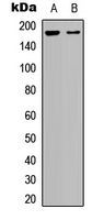
Western blot analysis of BAI1 expression in HepG2 (A); human brain (B) whole cell lysates.Application
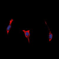
Immunofluorescent analysis of BAI1 staining in HepG2 cells. Formalin-fixed cells were permeabilized with 0.1% Triton X-100 in TBS for 5-10 minutes and blocked with 3% BSA-PBS for 30 minutes at room temperature. Cells were probed with the primary antibody in 3% BSA-PBS and incubated overnight at 4 i??i??C in a hidified chamber. Cells were washed with PBST and incubated with a DyLight 594-conjugated secondary antibody (red) in PBS at room temperature in the dark. DAPI was used to stain the cell nuclei (blue).| Product Name | BAI1 antibody |
|---|---|
| Antibody Type | Primary Antibodies |
| Product description | Rabbit Polyclonal antibody to BAI1Angiogenesis is controlled by a local balance between stimulators and inhibitors of new vessel growth and is suppressed under normal physiologic conditions. Angiogenesis has been shown to be essential for growth and metastasis of solid tumors. In order to obtain blood supply for their growth, tumor cells are potently angiogenic and attract new vessels as results of increased secretion of inducers and decreased production of endogenous negative regulators. BAI1 contains at least one 'functional' p53-binding site within an intron, and its expression has been shown to be induced by wildtype p53. There are two other brain-specific angiogenesis inhibitor genes, designated BAI2 and BAI3 which along with BAI1 have similar tissue specificities and structures, however only BAI1 is transcriptionally regulated by p53. BAI1 is postulated to be a member of the secretin receptor family, an inhibitor of angiogenesis and a growth suppressor of glioblastomas [provided by RefSeq, Jul 2008] |
| Immunogen | KLH-conjugated synthetic peptide encompassing a sequence within the center region of human BAI1. The exact sequence is proprietary. |
| Clonality | Polyclonal |
|---|---|
| Isotype | IgG |
| Host Species | Rabbit |
| Tested Applications | ICC/IFWB |
| WB: 1:500 - 1:1000. ICC/IF: 1:100 - 1:500: |
|
| Species Reactivity | Human |
| Concentration | 1mg/ml |
| Purification | Affinity purified |
| Gene Synonyms | ADGRB1 adhesion G protein-coupled receptor B1 GDAIF |
|---|---|
| Target | BAI1 |
| Tissue Specificity | Recognizes endogenous levels of BAI1 protein. |
Application
Western blot analysis of BAI1 expression in HepG2 (A); human brain (B) whole cell lysates.
Application
Immunofluorescent analysis of BAI1 staining in HepG2 cells. Formalin-fixed cells were permeabilized with 0.1% Triton X-100 in TBS for 5-10 minutes and blocked with 3% BSA-PBS for 30 minutes at room temperature. Cells were probed with the primary antibody in 3% BSA-PBS and incubated overnight at 4 i??i??C in a hidified chamber. Cells were washed with PBST and incubated with a DyLight 594-conjugated secondary antibody (red) in PBS at room temperature in the dark. DAPI was used to stain the cell nuclei (blue).| Application Notes | WB: 1:500 - 1:1000. ICC/IF: 1:100 - 1:500: |
|---|
| Form | Liquid |
|---|---|
| Storage Instructions | Shipped at 4 C. Upon delivery aliquot and store at -20 C for one year. Avoid freeze/thaw cycles. |
| Storage Buffer | Liquid in 0.42% Potassium phosphate, 0.87% Sodium chloride, pH 7.3, 30% glycerol, and 0.01% sodium azide. |


