Application
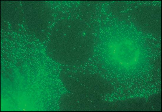
Immunofluorescent staining of 3T3 fibroblast with Monoclonal Anti-β1 and-β2 Adaptins (AP-1 & AP-2, Clone no. 100/1) .| Product Name | beta 1 + 2 Adaptin antibody |
|---|---|
| Antibody Type | Primary Antibodies |
| Product description | Mouse monoclonal [100 / 1] to beta 1 + 2 AdaptinCoated vesicle populations contain, besides clathrin, the adaptor complexes AP1 and AP2, also known as HAI (HA1) adaptor and HAII (HA2) adaptor or Assembly Protein 1 (AP1) and Assembly Protein 2 (AP2), respectively. Interest has focused on adaptors because they are known to mediate the interaction between clathrin and components of the membrane. Their location between the clathrin shell and the membrane, as revealed by cryoelectron microscopy, is consistent with such a function. In the cell, clathrin-adaptor interactions proceed in a very controlled and specific manner, leading to coated structures each containing only one type of adaptor. Clathrin-coated membranes at or originating from the plasma membrane contain the AP2 adaptor, while the AP1 adaptor appears to be largely restricted to clathrin-coated membranes of the trans-Golgi network. AP1 adaptor consists of four subunits termed beta 1 (formerly beta prime, 110 kDa), gamma (100 kDa), μ1 (47 kDa, and omega 1 (20 kDa). AP2 is made up of two 100-110 kDa polypeptides, termed alpha and beta 2 (formerly beta) subunits, μ2 (50 kDa), and s2 (17 kDa) subunit. Alpha and beta 2 subunits (alpha and beta 2 adaptins) behave in standard SDS-PAGE like 100-112 kDa polypeptides. Four alpha isoforms (alpha a1, alpha a2, alpha c1 and alpha c2) which are highly homologous in sequence have been described. ?1 and ?2 adaptins are highly homologous which suggests that the beta type adaptor chains may mediate the interaction of their adaptors with the highly conserved clathrin molecule. The alpha and gamma adaptins share only 25% overall identity. The other adaptor subunits of the two adaptor complexes which appear to be unrelated, are believed to possess binding sites for different cargo molecules (receptors) and for proteins which might regulate the complex functions of the adaptors. Monoclonal antibodies reacting specifically against adaptor proteins are useful tools for studies on the intracellular distribution and structural relationship of adaptor complexes. |
| Immunogen | AP2 adaptor from bovine brain. |
| Clonality | Monoclonal |
|---|---|
| Isotype | IgG1 |
| Clone Number | 100/1 |
| Host Species | Mouse |
| Tested Applications | ICC/IFIPWB |
| IF: Use at an assay dependent dilution. Stains clathrin-coated membranes at the plasma membrane, and in the Golgi region of methanol-fixed MDBK cells. WB: Use at a dilution of 1/200. This concentration was determined for bovine brain extract. With bovine and rat brain preparations, detects a major band at approximately 115 kDa and a minor component of approximately 110 kDa of AP1, and bands at 104 or 106 and 112 kDa of AP2. The antibody reacts with polypeptides of approximately 100 kDa in bovine liver, human heart fibroblasts and Madin-Darby bovine kidney cultured cells (MDBK), but not with any components in the 110-115 kDa range from these sources, or from rat pheochromocytoma cultured cells (PC12), neuroblastoma or astrocytes. Optimal dilutions/concentrations should be determined by the end user.: |
|
| Species Reactivity | HumanRatBovine |
| Concentration | 1mg/ml |
| Purification | Affinity purified |
| Gene Synonyms | CLAPB2 DKFZp781K0743 162 600157 AP105B ADTB1 beta 1 + 2 Adaptin ADTB2 CLAPB1 AP2BETA BAM22 AP2B1 601025 AP105A AP2-BETA 163 beta1 + 2 Adaptin P63010 AP1B1 Q10567 MGC128441 AP2 BETA |
|---|---|
| Target | beta 1 + 2 Adaptin |
| Cellular Localization | Cytoplasmic |
Application
Immunofluorescent staining of 3T3 fibroblast with Monoclonal Anti-β1 and-β2 Adaptins (AP-1 & AP-2, Clone no. 100/1) .| Application Notes | IF: Use at an assay dependent dilution. Stains clathrin-coated membranes at the plasma membrane, and in the Golgi region of methanol-fixed MDBK cells. WB: Use at a dilution of 1/200. This concentration was determined for bovine brain extract. With bovine and rat brain preparations, detects a major band at approximately 115 kDa and a minor component of approximately 110 kDa of AP1, and bands at 104 or 106 and 112 kDa of AP2. The antibody reacts with polypeptides of approximately 100 kDa in bovine liver, human heart fibroblasts and Madin-Darby bovine kidney cultured cells (MDBK), but not with any components in the 110-115 kDa range from these sources, or from rat pheochromocytoma cultured cells (PC12), neuroblastoma or astrocytes. Optimal dilutions/concentrations should be determined by the end user.: |
|---|
| Form | Liquid |
|---|---|
| Storage Instructions | Keep as concentrated solution. Store at 4ºC short term. For extended storage aliquot and store at -20ºC or below. Avoid freeze-thaw cycles. |
| Storage Buffer | Ascites fluid containing 0.1% Sodium azide |


