Application
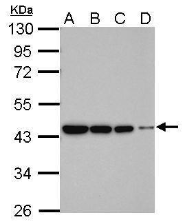
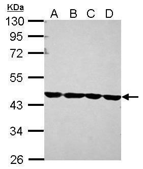
Sample (whole cell lysate) A: 293T 20ug B: 293T 10ug C: 293T 5ug D: 293T 1ug 10% SDS PAGE diluted at 1:5000 The HRP-conjugated anti-rabbit IgG antibody was used to detect the primary antibody.Application
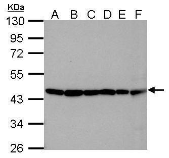
Sample (30 µg of whole cell lysate) A: 293T B: NTH-3T3 C: mouse brain D: PC-12 E: Rat brain F: drosophila 10% SDS PAGE diluted at 1:10000 The HRP-conjugated anti-rabbit IgG antibody was used to detect the primary antibody.Application
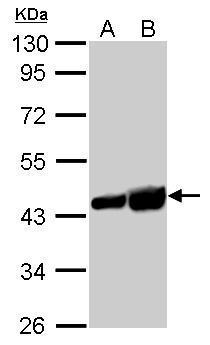
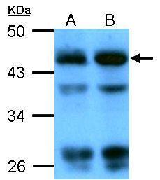
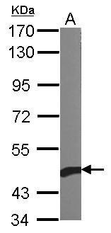
beta Actin antibody detects ACTB protein by western blot analysis. A. 30 µg PC-12 whole cell lysate/extract B. 30 µg Rat-2 whole cell lysate/extract 10% SDS-PAGE beta Actin antibody dilution: 1:5000 The HRP-conjugated anti-rabbit IgG antibody was used to detect the primary antibody.Application
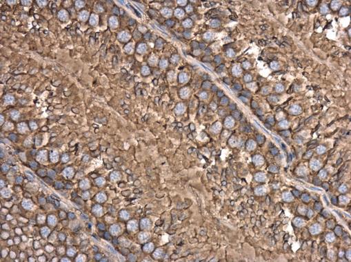
beta Actin antibody detects beta Actin protein at cytoplasm in rat testis by immunohistochemical analysis. Sample: Paraffin-embedded rat testis. beta Actin antibody diluted at 1:500.Application
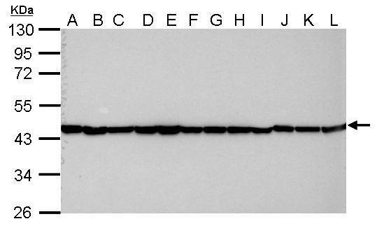
Sample (30 µg of whole cell lysate) A: Jurkat B: Raji C: 293T D: A431 E: HeLa F: HepG2 G: H1299 H: HCT116 I: MCF-7 J: NT2D1 K: PC-3 L: U87-MG 10% SDS PAGE diluted at 1:10000 The HRP-conjugated anti-rabbit IgG antibody was used to detect the primary antibody.Application
Sample (whole cell lysate) A: Candida albicans (sc5314) 20ug B: Candida albicans (sc5314) 40ug 10% SDS PAGE diluted at 1:4000 The HRP-conjugated anti-rabbit IgG antibody was used to detect the primary antibody.Application
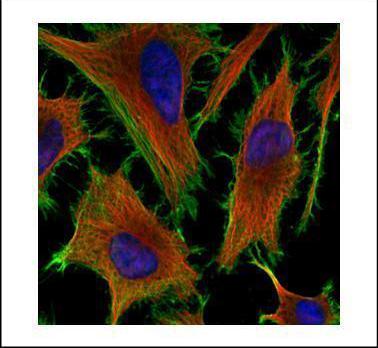
Confocal immunofluorescence analysis (Olympus FV10i) of methanol-fixed HeLa, using beta Actin antibody (Green) at 1:200 dilution. Alpha-tubulin filaments were labeled with (Red) at 1:2000.Application
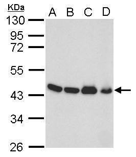
beta Actin antibody detects ACTB protein by western blot analysis. A. 50 µg mouse brain lysate/extract B. 50 µg mouse kidney lysate/extract C. 50 µg mouse lung lysate/extract D. 50 µg mouse testis lysate/extract 10% SDS-PAGE beta Actin antibody dilution: 1:10000 The HRP-conjugated anti-rabbit IgG antibody was used to detect the primary antibody.Application
Sample (30 µg of whole cell lysate) A: 293T B: A431 C: Jurkat D: Raji 10% SDS PAGE diluted at 1:1000 The HRP-conjugated anti-rabbit IgG antibody was used to detect the primary antibody.Application
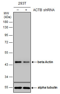
Non-transfected (–) and transfected (+) 293T whole cell extracts (10 µg) were separated by 10% SDS-PAGE, and the membrane was blotted with beta Actin antibody diluted at 1:15000. The HRP-conjugated anti-rabbit IgG antibody was used to detect the primary antibody.Application
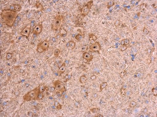
beta Actin antibody detects beta Actin protein at cytoplasm in mouse brain by immunohistochemical analysis. Sample: Paraffin-embedded mouse brain. beta Actin antibody diluted at 1:500.Application
Sample (10 µg of whole cell lysate) A: Yeast lysate 7.5% SDS PAGE diluted at 1:1000 The HRP-conjugated anti-rabbit IgG antibody was used to detect the primary antibody.Application
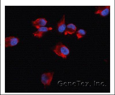
Immunofluorescence analysis of methanol-fixed HeLa, using Beta-actin antibody at 1:500 dilution.Application
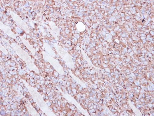
Immunohistochemical analysis of paraffin-embedded H1299 xenograft, using Beta actin antibody at 1:100 dilution.WB
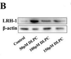
(B) Western blotting of LRH-1 protein in granulosa cells by treatment with various concentrations of DLPC.WB
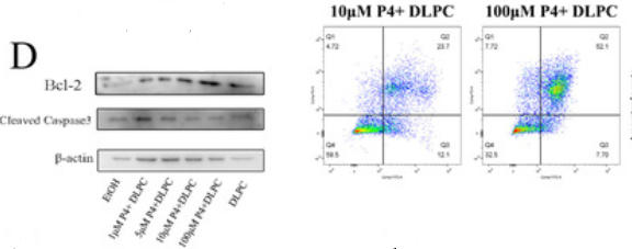
(D) Western blotting of Bcl2 and cleaved-caspase 3 in granulosa cells by treatment with 50 μM DLPC, 50 μM DLPC + 1 μM P4, 50 μM DLPC + 5 μM P4, 50 μM DLPC + 10 μM P4 and 50 μM DLPC + 100 μM P4, respectively| Product Name | beta Actin antibody |
|---|---|
| Antibody Type | Primary Antibodies |
| Product description | Rabbit Polyclonal antibody to beta Actin (actin, beta)This gene encodes one of six different actin proteins. Actins are highly conserved proteins that are involved in cell motility, structure, and integrity. This actin is a major constituent of the contractile apparatus and one of the two nonmuscle cytoskeletal actins. [provided by RefSeq] |
| Immunogen | Carrier-protein conjugated synthetic peptide encompassing a sequence within the C-terminus region of human beta Actin. The exact sequence is proprietary. |
| Clonality | Polyclonal |
|---|---|
| Isotype | IgG |
| Host Species | Rabbit |
| Tested Applications | IBICC/IFIHC-PIPWB |
| *Optimal dilutions/concentrations should be determined by the researcher. | |
| Species Reactivity | Candida albicansCatDrosophilaHumanMouseRatRiceYeast |
| Predicted Reactivity | Japanese medaka, Xenopus laevis, Dog, Rabbit, Chicken, Sheep, Rhesus Monkey, Chimpanzee, Bovine, Guinea pigJapanese medaka (98%), Xenopus laevis (99%), Dog (100%), Rabbit (99%), Chicken (100%), Sheep (100%), Rhesus Monkey (100%), Chimpanzee (100%), Bovine (100%), Guinea pig (100%) |
| Concentration | 0.2mg/ml |
| Purification | Affinity purified |
| Gene Synonyms | PS1TP5BP1 antibody, ACTB antibody, actin, cytoplasmic 1 antibody, beta cytoskeletal actin antibody, PS1TP5-binding protein 1 antibody, actin, beta antibody |
|---|---|
| Molecular Weight(MW) | 42 kDa (note) |
| Cellular Localization | Cytoplasm , cytoskeleton |
Application
Sample (whole cell lysate) A: 293T 20ug B: 293T 10ug C: 293T 5ug D: 293T 1ug 10% SDS PAGE diluted at 1:5000 The HRP-conjugated anti-rabbit IgG antibody was used to detect the primary antibody.
Application
Sample (30 µg of whole cell lysate) A: 293T B: NTH-3T3 C: mouse brain D: PC-12 E: Rat brain F: drosophila 10% SDS PAGE diluted at 1:10000 The HRP-conjugated anti-rabbit IgG antibody was used to detect the primary antibody.
Application
beta Actin antibody detects ACTB protein by western blot analysis. A. 30 µg PC-12 whole cell lysate/extract B. 30 µg Rat-2 whole cell lysate/extract 10% SDS-PAGE beta Actin antibody dilution: 1:5000 The HRP-conjugated anti-rabbit IgG antibody was used to detect the primary antibody.
Application
beta Actin antibody detects beta Actin protein at cytoplasm in rat testis by immunohistochemical analysis. Sample: Paraffin-embedded rat testis. beta Actin antibody diluted at 1:500.
Application
Sample (30 µg of whole cell lysate) A: Jurkat B: Raji C: 293T D: A431 E: HeLa F: HepG2 G: H1299 H: HCT116 I: MCF-7 J: NT2D1 K: PC-3 L: U87-MG 10% SDS PAGE diluted at 1:10000 The HRP-conjugated anti-rabbit IgG antibody was used to detect the primary antibody.
Application
Sample (whole cell lysate) A: Candida albicans (sc5314) 20ug B: Candida albicans (sc5314) 40ug 10% SDS PAGE diluted at 1:4000 The HRP-conjugated anti-rabbit IgG antibody was used to detect the primary antibody.
Application
Confocal immunofluorescence analysis (Olympus FV10i) of methanol-fixed HeLa, using beta Actin antibody (Green) at 1:200 dilution. Alpha-tubulin filaments were labeled with (Red) at 1:2000.
Application
beta Actin antibody detects ACTB protein by western blot analysis. A. 50 µg mouse brain lysate/extract B. 50 µg mouse kidney lysate/extract C. 50 µg mouse lung lysate/extract D. 50 µg mouse testis lysate/extract 10% SDS-PAGE beta Actin antibody dilution: 1:10000 The HRP-conjugated anti-rabbit IgG antibody was used to detect the primary antibody.
Application
Sample (30 µg of whole cell lysate) A: 293T B: A431 C: Jurkat D: Raji 10% SDS PAGE diluted at 1:1000 The HRP-conjugated anti-rabbit IgG antibody was used to detect the primary antibody.
Application
Non-transfected (–) and transfected (+) 293T whole cell extracts (10 µg) were separated by 10% SDS-PAGE, and the membrane was blotted with beta Actin antibody diluted at 1:15000. The HRP-conjugated anti-rabbit IgG antibody was used to detect the primary antibody.
Application
beta Actin antibody detects beta Actin protein at cytoplasm in mouse brain by immunohistochemical analysis. Sample: Paraffin-embedded mouse brain. beta Actin antibody diluted at 1:500.
Application
Sample (10 µg of whole cell lysate) A: Yeast lysate 7.5% SDS PAGE diluted at 1:1000 The HRP-conjugated anti-rabbit IgG antibody was used to detect the primary antibody.
Application
Immunofluorescence analysis of methanol-fixed HeLa, using Beta-actin antibody at 1:500 dilution.
Application
Immunohistochemical analysis of paraffin-embedded H1299 xenograft, using Beta actin antibody at 1:100 dilution.
WB
(B) Western blotting of LRH-1 protein in granulosa cells by treatment with various concentrations of DLPC.
WB
(D) Western blotting of Bcl2 and cleaved-caspase 3 in granulosa cells by treatment with 50 μM DLPC, 50 μM DLPC + 1 μM P4, 50 μM DLPC + 5 μM P4, 50 μM DLPC + 10 μM P4 and 50 μM DLPC + 100 μM P4, respectively| Positive Control | 293T , A341 , Jurkat , Raji , HeLa , HepG2 , H1299 , HCT116 , MCF-7 , NT2D1 , PC-3 , U87-MG , NIH-3T3 , mouse brain , PC-12 , Rat2 , Rat brain , drosophila , Yeast lysate , Candida albicans (sc5314) whole cell lysate , *Rice leaf , *mouse aorta , *H9C2 , *OKT3 , *mouse ileum tissue , *mouse heart tissue , *mouse skeletal muscle tissue , *mouse brain tissue , *human periodontal ligament |
|---|---|
| Application Notes | *Optimal dilutions/concentrations should be determined by the researcher. |
| Form | Liquid |
|---|---|
| Storage Instructions | Keep as concentrated solution. Aliquot and store at -20ºC or below. Avoid multiple freeze-thaw cycles. |
| Storage Buffer | 1XPBS, 1% BSA, 20% Glycerol (pH7). 0.025% ProClin 300 was added as a preservative. |


