Application
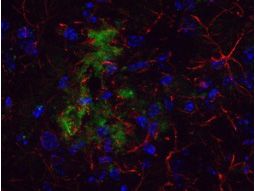
Immunohistochemistical detection of beta Amyloid using Anti-Beta Amyloid Antibody on TG APP23 mouse brain cortex frozen sections. Anti-Beta Amyloid Antibody used at 1/200 and incubated for 2 hours in TBS/BSA/Tween/azide. Fluorescent labelled anti rabbit IgG was then added.Application
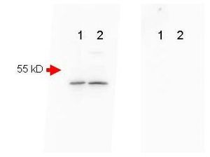
Mouse Brain (Lane 1) and Mouse Spinal Chord (Lane 2) were run on a 4-20% gradient gel, Blocked in 1% BSA-TBS-T 30 min RT and probed with Rb-a-Beta Amyloid 1:1000 in 1% BSA-TBS-T o/n 4°C. FEMTOMAX chemiluminescent substrate was used for detection of a 40-50 kD band consistent with a higher MW precursor form of beta amyloid. A secondary Ab only control (Shown right) showed no detectable signal.Application
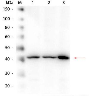
Western Blot of Rabbit anti-Beta Amyloid Antibody. Lane 1: HEK293 WCL. Lane 2: Mouse Brain WCL. Lane 3: A-172 WCL. Load: 10.0 µg per lane. Primary antibody: Beta Amyloid Antibody at 1:1,000 overnight at 4°C. Secondary antibody: Peroxidase Conjugated Goat-a-Rabbit IgG at 1:40,000 for 30 min at RT. Block for 30 min at RT. Predicted/Observed size: 40 kDa, 40 kDa for Beta Amyloid.Application
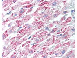
Human Heart (formalin-fixed, paraffin-embedded) stained with Anti-Beta Amyloid Antibody at 5 ug/ml followed by biotinylated goat anti-rabbit IgG secondary antibody, alkaline phosphatase-streptavidin and chromogen.| Product Name | beta Amyloid antibody |
|---|---|
| Antibody Type | Primary Antibodies |
| Product description | Rabbit Polyclonal antibody to beta AmyloidBeta Amyloid, often abbreviated as A-beta, is a protein that builds up in the brains of persons with Alzheimer's disease, collecting in clumps called plaques or senile plaques. While some researchers question whether Beta Amyloid is the cause of the dementia, most agree that it is involved in the disruption of thinking that is a hallmark of the disease. In some cases of familial Alzheimer's disease, mutations in genes for the proteins called the presenilins lead to increased production of myloid. Researchers have been looking at how presenilin-1 in particular contributes to the excess buildup of Beta Amyloid. Presenilin-1 apparently acts to increase the activity of gamma-secretase, an enzyme that changes a normal protein (amyloid precursor protein or APP) into Beta Amyloid itself. Furthermore, presenilin-1 might be gamma-secretase. |
| Immunogen | Peptide - amino acids 1-14 of human beta amyloid |
| Clonality | Polyclonal |
|---|---|
| Isotype | IgG |
| Host Species | Rabbit |
| Tested Applications | ELISAICC/IFIHC-FIHC-PWB |
| *Optimal dilutions/concentrations should be determined by the researcher. | |
| Species Reactivity | HumanMouse |
| Concentration | 1mg/ml (Please refer to the vial label for the specific concentration) |
| Purification | Affinity purified |
| Gene Synonyms | PN2 , AAA , CVAP , ABPP , AD1 , APP , APPI , CTFGAMMA , PN II , ABETA |
|---|---|
| Target | beta Amyloid |
Application
Immunohistochemistical detection of beta Amyloid using Anti-Beta Amyloid Antibody on TG APP23 mouse brain cortex frozen sections. Anti-Beta Amyloid Antibody used at 1/200 and incubated for 2 hours in TBS/BSA/Tween/azide. Fluorescent labelled anti rabbit IgG was then added.
Application
Mouse Brain (Lane 1) and Mouse Spinal Chord (Lane 2) were run on a 4-20% gradient gel, Blocked in 1% BSA-TBS-T 30 min RT and probed with Rb-a-Beta Amyloid 1:1000 in 1% BSA-TBS-T o/n 4°C. FEMTOMAX chemiluminescent substrate was used for detection of a 40-50 kD band consistent with a higher MW precursor form of beta amyloid. A secondary Ab only control (Shown right) showed no detectable signal.
Application
Western Blot of Rabbit anti-Beta Amyloid Antibody. Lane 1: HEK293 WCL. Lane 2: Mouse Brain WCL. Lane 3: A-172 WCL. Load: 10.0 µg per lane. Primary antibody: Beta Amyloid Antibody at 1:1,000 overnight at 4°C. Secondary antibody: Peroxidase Conjugated Goat-a-Rabbit IgG at 1:40,000 for 30 min at RT. Block for 30 min at RT. Predicted/Observed size: 40 kDa, 40 kDa for Beta Amyloid.
Application
Human Heart (formalin-fixed, paraffin-embedded) stained with Anti-Beta Amyloid Antibody at 5 ug/ml followed by biotinylated goat anti-rabbit IgG secondary antibody, alkaline phosphatase-streptavidin and chromogen.| Application Notes | *Optimal dilutions/concentrations should be determined by the researcher. |
|---|
| Form | Liquid |
|---|---|
| Storage Instructions | Store vial at -20° C prior to opening. Aliquot contents and freeze at -20° C or below for extended storage. Avoid cycles of freezing and thawing. |
| Storage Buffer | 0.02 M Potassium Phosphate, 0.15 M Sodium Chloride, pH 7.2 and 0.01% (w/v) Sodium Azide |


