Application
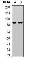
Western blot analysis of Beta-catenin (pS33) expression in HeLa (A); HepG2 (B) whole cell lysates.Application
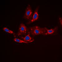
Immunofluorescent analysis of Beta-catenin (pS33) staining in HepG2 cells. Formalin-fixed cells were permeabilized with 0.1% Triton X-100 in TBS for 5-10 minutes and blocked with 3% BSA-PBS for 30 minutes at room temperature. Cells were probed with the primary antibody in 3% BSA-PBS and incubated overnight at 4 deg C in a hidified chamber. Cells were washed with PBST and incubated with a DyLight 594-conjugated secondary antibody (red) in PBS at room temperature in the dark. DAPI was used to stain the cell nuclei (blue).Application
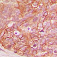
Immunohistochemical analysis of Beta-catenin (pS33) staining in human breast cancer formalin fixed paraffin embedded tissue section. The section was pre-treated using heat mediated antigen retrieval with sodium citrate buffer (pH 6.0). The section was then incubated with the antibody at room temperature and detected using an HRP conjugad compact polymer system. DAB was used as the chromogen. The section was then counterstained with haematoxylin and mounted with DPX.| Product Name | beta Catenin (phospho Ser33) antibody |
|---|---|
| Antibody Type | Primary Antibodies |
| Product description | Rabbit Polyclonal antibody to beta Catenin (phospho Ser33)The protein encoded by this gene is part of a complex of proteins that constitute adherens junctions (AJs). AJs are necessary for the creation and maintenance of epithelial cell layers by regulating cell growth and adhesion between cells. The encoded protein also anchors the actin cytoskeleton and may be responsible for transmitting the contact inhibition signal that causes cells to stop dividing once the epithelial sheet is complete. Finally, this protein binds to the product of the APC gene, which is mutated in adenomatous polyposis of the colon. Mutations in this gene are a cause of colorectal cancer (CRC), pilomatrixoma (PTR), medulloblastoma (MDB), and ovarian cancer. Three transcript variants encoding the same protein have been found for this gene.[provided by RefSeq, Oct 2009] |
| Immunogen | KLH-conjugated synthetic peptide encompassing a sequence within the N-term region of human Beta-catenin (pS33). The exact sequence is proprietary. |
| Modification | p-Ser33 |
| Clonality | Polyclonal |
|---|---|
| Isotype | IgG |
| Host Species | Rabbit |
| Tested Applications | ICC/IFIHC-PWB |
| WB: 1:500 - 1:1000. IHC-P: 1:100 - 1:200. ICC/IF: 1:100 - 1:500 | |
| Species Reactivity | Human |
| Concentration | 1mg/ml |
| Purification | Affinity purified |
| Gene Synonyms | armadillo , catenin (cadherin-associated protein), beta 1, 88kDa , MRD19 , CTNNB , CTNNB1 |
|---|---|
| Target | beta Catenin (phospho Ser33) |
| Tissue Specificity | Recognizes endogenous levels of Beta-catenin (pS33) protein. |
Application
Western blot analysis of Beta-catenin (pS33) expression in HeLa (A); HepG2 (B) whole cell lysates.
Application
Immunofluorescent analysis of Beta-catenin (pS33) staining in HepG2 cells. Formalin-fixed cells were permeabilized with 0.1% Triton X-100 in TBS for 5-10 minutes and blocked with 3% BSA-PBS for 30 minutes at room temperature. Cells were probed with the primary antibody in 3% BSA-PBS and incubated overnight at 4 deg C in a hidified chamber. Cells were washed with PBST and incubated with a DyLight 594-conjugated secondary antibody (red) in PBS at room temperature in the dark. DAPI was used to stain the cell nuclei (blue).
Application
Immunohistochemical analysis of Beta-catenin (pS33) staining in human breast cancer formalin fixed paraffin embedded tissue section. The section was pre-treated using heat mediated antigen retrieval with sodium citrate buffer (pH 6.0). The section was then incubated with the antibody at room temperature and detected using an HRP conjugad compact polymer system. DAB was used as the chromogen. The section was then counterstained with haematoxylin and mounted with DPX.| Application Notes | WB: 1:500 - 1:1000. IHC-P: 1:100 - 1:200. ICC/IF: 1:100 - 1:500 |
|---|
| Form | Liquid |
|---|---|
| Storage Instructions | Shipped at 4 °C. Upon delivery aliquot and store at -20 °C for one year. Avoid freeze/thaw cycles. |
| Storage Buffer | Liquid in 0.42% Potassium phosphate; 0.87% Sodium chloride; pH 7.3; 30% glycerol; and 0.01% sodium azide. |


