Application
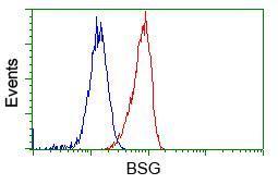
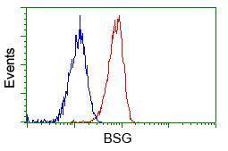
Flow cytometric Analysis of Hela cells, using anti-BSG antibody,(Red), compared to a nonspecific negative control antibody(TA50011),(Blue).Application
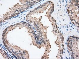
Immunohistochemical staining of paraffin-embedded Human prostate tissue using anti-BSG mouse monoclonal antibody. ( Dilution 1:50)Application
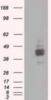
HEK293T cells were transfected with the pCMV6-ENTRY control (Left lane) or pCMV6-ENTRY BSG (Right lane) cDNA for 48 hrs and lysed. Equivalent amounts of cell lysates (5 ug per lane) were separated by SDS-PAGE and immunoblotted with anti-BSG.Application
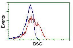
HEK293T cells transfected with either RC219464 overexpress plasmid(Red) or empty vector control plasmid(Blue) were immunostained by anti-BSG antibody, and then analyzed by flow cytometry.Application
Flow cytometric Analysis of Jurkat cells, using anti-BSG antibody,(Red), compared to a nonspecific negative control antibody(TA50011),(Blue).Application
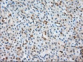
Immunohistochemical staining of paraffin-embedded Human pancreas tissue using anti-BSG mouse monoclonal antibody. ( Dilution 1:50)Application
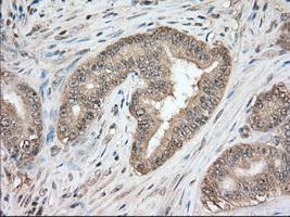
Immunohistochemical staining of paraffin-embedded Adenocarcinoma of Human colon tissue using anti-BSG mouse monoclonal antibody. ( Dilution 1:50)Application
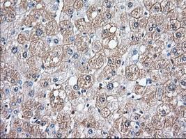
Immunohistochemical staining of paraffin-embedded Human liver tissue using anti-BSG mouse monoclonal antibody. ( Dilution 1:50)Application
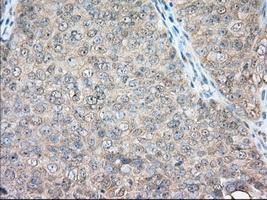
Immunohistochemical staining of paraffin-embedded Adenocarcinoma of Human ovary tissue using anti-BSG mouse monoclonal antibody. ( Dilution 1:50)Application
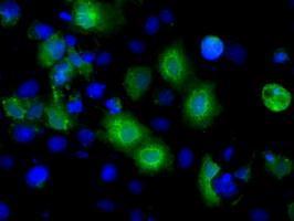
Anti-BSG mouse monoclonal antibody immunofluorescent staining of COS7 cells transiently transfected with BSG| Product Name | BSG antibody [5D8] |
|---|---|
| Antibody Type | Primary Antibodies |
| Product description | Mouse monoclonal antibody [5D8] to BSGPlays pivotal roles in spermatogenesis, embryo implantation, neural network formation and tumor progression. Stimulates adjacent fibroblasts to produce matrix metalloproteinases (MMPS). May target monocarboxylate transporters SLC16A1, SLC16A3 and SLC16A8 to plasma membranes of retinal pigment epithelium and neural retina. Seems to be a receptor for oligomannosidic glycans. In vitro, promotes outgrowth of astrocytic processes. |
| Immunogen | Full length human recombinant protein of human BSG(NP_001719) produced in HEK293T cell. |
| Clonality | Monoclonal |
|---|---|
| Isotype | IgG2 |
| Clone Number | 5D8 |
| Host Species | Mouse |
| Tested Applications | FACS,ICC/IF,IHC,IHC-P,WB |
| IHC 1:50 | |
| Species Reactivity | Human |
| Concentration | 1mg/ml |
| Purification | Affinity purified |
| Gene Synonyms | EMMPRIN, OK, TCSF, BSG, 5F7, M6, CD147 |
|---|---|
| Target | BSG |
| Tissue Specificity | is specific for human BSG(NP_001719). |
| Cellular Localization | Colocalizes with SLC16A1 and SLC16A8 (By similarity). Identified by mass spectrometry in melanosome fractions from stage I to stage IV. |
Application
Flow cytometric Analysis of Hela cells, using anti-BSG antibody,(Red), compared to a nonspecific negative control antibody(TA50011),(Blue).
Application
Immunohistochemical staining of paraffin-embedded Human prostate tissue using anti-BSG mouse monoclonal antibody. ( Dilution 1:50)
Application
HEK293T cells were transfected with the pCMV6-ENTRY control (Left lane) or pCMV6-ENTRY BSG (Right lane) cDNA for 48 hrs and lysed. Equivalent amounts of cell lysates (5 ug per lane) were separated by SDS-PAGE and immunoblotted with anti-BSG.
Application
HEK293T cells transfected with either RC219464 overexpress plasmid(Red) or empty vector control plasmid(Blue) were immunostained by anti-BSG antibody, and then analyzed by flow cytometry.
Application
Flow cytometric Analysis of Jurkat cells, using anti-BSG antibody,(Red), compared to a nonspecific negative control antibody(TA50011),(Blue).
Application
Immunohistochemical staining of paraffin-embedded Human pancreas tissue using anti-BSG mouse monoclonal antibody. ( Dilution 1:50)
Application
Immunohistochemical staining of paraffin-embedded Adenocarcinoma of Human colon tissue using anti-BSG mouse monoclonal antibody. ( Dilution 1:50)
Application
Immunohistochemical staining of paraffin-embedded Human liver tissue using anti-BSG mouse monoclonal antibody. ( Dilution 1:50)
Application
Immunohistochemical staining of paraffin-embedded Adenocarcinoma of Human ovary tissue using anti-BSG mouse monoclonal antibody. ( Dilution 1:50)
Application
Anti-BSG mouse monoclonal antibody immunofluorescent staining of COS7 cells transiently transfected with BSG| Application Notes | IHC 1:50 |
|---|
| Form | Liquid |
|---|---|
| Storage Instructions | For short-term storage, store at 4°C or aliquot into working amounts and store at -20°C. For long-term storage, store at -70°C (aliquotted). Avoid repeated freeze-thaw cycles. |
| Storage Buffer | PBS (PH 7.3) containing 1% BSA, 50% glycerol and 0.02% sodium azide. |


