Application
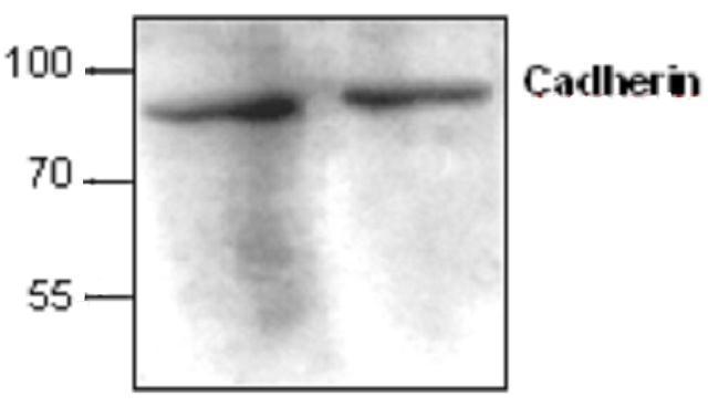
Western blots analysis of Cadherin expression with Jurkat cell lysates.| Product Name | Cadherin antibody |
|---|---|
| Antibody Type | Primary Antibodies |
| Product description | Rabbit polyclonal antibody to CadherinCadherins comprise a family of Ca-dependent adhesion molecules that function to mediate cell-cell binding critical to the maintenance of tissue structure and morphogenesis. Cadherins consist of large extracellular domains characterized by a series of five homologous NH2 terminal repeats. The most distal of cadherins is though to be responsible for binding specificity, transmembrane domains and carboxy terminal domains. The relative short intracellular domains interact with a variety of cytoplasmic proteins, such as beta-catenin, to regulate cadherin function. |
| Clonality | Polyclonal |
|---|---|
| Isotype | IgG |
| Host Species | Rabbit |
| Tested Applications | IHCIPWB |
| Western blotting (1-2 µg/ml), immunoprecipitation (10-20 µg/ml), and Immunohistochemistry (10-20 µg/ml). However, the optimal conditions should be determined individually. The antibody detects E- and P-cadherin (~95 kDa). Jurkat cell lysate can be used as a positive control.: |
|
| Species Reactivity | HumanMouse |
| Concentration | 0.5mg/ml |
| Purification | Affinity purified |
| Target | Cadherin |
|---|---|
| Tissue Specificity | This antibody is specific for Cadherin. |
| Cellular Localization | Colocalizes with DLGAP5 at sites of cell-cell contact in intestinal epithelial cells. Anchored to actin microfilaments through association with alpha-, beta- and gamma-catenin. Sequential proteolysis induced by apoptosis or calcium influx, results in tran |
Application
Western blots analysis of Cadherin expression with Jurkat cell lysates.| Application Notes | Western blotting (1-2 µg/ml), immunoprecipitation (10-20 µg/ml), and Immunohistochemistry (10-20 µg/ml). However, the optimal conditions should be determined individually. The antibody detects E- and P-cadherin (~95 kDa). Jurkat cell lysate can be used as a positive control.: |
|---|
| Form | Liquid |
|---|---|
| Storage Instructions | The antibody solution should be gently mixed before use. Stored at -20 °C for future use. |
| Storage Buffer | PBS (pH 7.2) containing 30% glycerol, 0.5% BSA, 0.01% thimerosal |


