Application
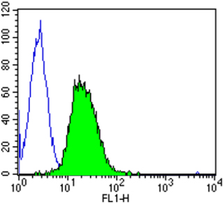
Flow cytometry analysis of Calmodulin showing positive staining in the cytoplasm of C6 cells compared to an isotype control (blue). Cells were harvested, adjusted to a concentration of 1-5x10^6 cells/ml, fixed with 2% paraformaldehyde and washed with PBS. Cells were penetrated by dropping the supernatant, adding 90% methanol and incubated for 10 minutes at room temperature. Follwing penetration, cells were blocked with a 2% solution of BSA-PBS for 30 min at room temperature and incubated with a Calmodulin monoclonal antibody at a dilution of 2 ug/test for 60 min at room temperature. Cells were then incubated for 40 min at room temperature in the dark using a Dylight 488-conjugated goat anti-mouse IgG (H+L) secondary antibody and re-suspended in PBS for FACS analysis.Application
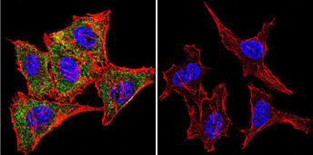
Immunofluorescent analysis of Calmodulin using Calmodulin Monoclonal antibody (2D1) shows staining in HeLa cells. Calmodulin staining (green), F-Actin staining with Phalloidin (red) and nuclei with DAPI (blue) is shown. Cells were grown on chamber slides and fixed with formaldehyde prior to staining. Cells were probed without (control) or with or an antibody recognizing Calmodulin at a dilution of 1:20 over night at 4 degrees C, washed with PBS and incubated with a DyLight-488 conjugated secondary antibody.Application
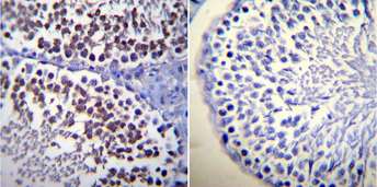
Immunohistochemistry was performed on normal biopsies of deparaffinized Rat testis tissue. To expose target proteins, heat induced antigen retrieval was performed using 10mM sodium citrate (pH6.0) buffer, microwaved for 8-15 minutes. Following antigen retrieval tissues were blocked in 3% BSA-PBS for 30 minutes at room temperature. Tissues were then probed at a dilution of 1:20 with a mouse monoclonal antibody recognizing Calmodulin or without primary antibody (negative control) overnight at 4°C in a humidified chamber. Tissues were washed extensively with PBST and endogenous peroxidase activity was quenched with a peroxidase suppressor. Detection was performed using a biotin-conjugated secondary antibody and SA-HRP, followed by colorimetric detection using DAB. Tissues were counterstained with hematoxylin and prepped for mounting.Application
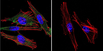
Immunofluorescent analysis of Calmodulin using Calmodulin Monoclonal antibody (2D1) shows staining in A2058 melanoma cells. Calmodulin staining (green), F-Actin staining with Phalloidin (red) and nuclei with DAPI (blue) is shown. Cells were grown on chamber slides and fixed with formaldehyde prior to staining. Cells were probed without (control) or with or an antibody recognizing Calmodulin at a dilution of 1:20 over night at 4 degrees C, washed with PBS and incubated with a DyLight-488 conjugated secondary antibody.Application
Immunofluorescent analysis of Calmodulin using Calmodulin Monoclonal antibody (2D1) shows staining in C6 glioma cells. Calmodulin staining (green), F-Actin staining with Phalloidin (red) and nuclei with DAPI (blue) is shown. Cells were grown on chamber slides and fixed with formaldehyde prior to staining. Cells were probed without (control) or with or an antibody recognizing Calmodulin at a dilution of 1:20 over night at 4 degrees C, washed with PBS and incubated with a DyLight-488 conjugated secondary antibody.Application
Flow cytometry analysis of Calmodulin showing positive staining in the cytoplasm of PC12 cells compared to an isotype control (blue). Cells were harvested, adjusted to a concentration of 1-5x10^6 cells/ml, fixed with 2% paraformaldehyde and washed with PBS. Cells were penetrated by dropping the supernatant, adding 90% methanol and incubated for 10 minutes at room temperature. Follwing penetration, cells were blocked with a 2% solution of BSA-PBS for 30 min at room temperature and incubated with a Calmodulin monoclonal antibody at a dilution of 2 ug/test for 60 min at room temperature. Cells were then incubated for 40 min at room temperature in the dark using a Dylight 488-conjugated goat anti-mouse IgG (H+L) secondary antibody and re-suspended in PBS for FACS analysis.Application
Western blot of purified calmodulin usingApplication
Flow cytometry analysis of Calmodulin showing positive staining in the cytoplasm of MCF-7 cells compared to an isotype control (blue). Cells were harvested, adjusted to a concentration of 1-5x10^6 cells/ml, fixed with 2% paraformaldehyde and washed with PBS. Cells were penetrated by dropping the supernatant, adding 90% methanol and incubated for 10 minutes at room temperature. Follwing penetration, cells were blocked with a 2% solution of BSA-PBS for 30 min at room temperature and incubated with a Calmodulin monoclonal antibody at a dilution of 2 ug/test for 60 min at room temperature. Cells were then incubated for 40 min at room temperature in the dark using a Dylight 488-conjugated goat anti-mouse IgG (H+L) secondary antibody and re-suspended in PBS for FACS analysis.Application
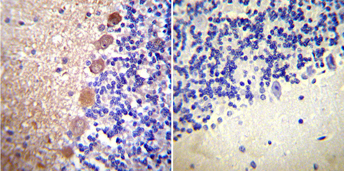
Immunohistochemistry was performed on normal biopsies of de-paraffinized Rat cerebellum tissue. To expose target proteins, heat induced antigen retrieval was performed using 10mM sodium citrate (pH6.0) buffer, microwaved for 8-15 minutes. Following antigen retrieval tissues were blocked in 3% BSA-PBS for 30 minutes at room temperature. Tissues were then probed at a dilution of 1:20 with a mouse monoclonal antibody recognizing Calmodulin or without primary antibody (negative control) overnight at 4°C in a humidified chamber. Tissues were washed extensively with PBST and endogenous peroxidase activity was quenched with a peroxidase suppressor. Detection was performed using a biotin-conjugated secondary antibody and SA-HRP, followed by colorimetric detection using DAB. Tissues were counterstained with hematoxylin and prepped for mounting.Application
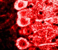
Immunolocalization of calmodulin in rat brain using| Product Name | Calmodulin antibody |
|---|---|
| Antibody Type | Primary Antibodies |
| Product description | Mouse Monoclonal antibody to CalmodulinCalmodulin is a small, highly conserved calcium binding protein found in all eukaryotic cells. With the capacity to bind up to four calcium ions, this 17 kDa protein acts as an important intracellular receptor for regulatory calcium signals. As it binds calcium, calmodulin undergoes conformational changes which can increase its affinity for target proteins. It acts both directly, through interaction with key target enzymes, and indirectly, via specific kinases. Studies have found that calmodulin participates in the regulation of several biological processes including energy and biosynthetic metabolism, cell motility, exocytosis, cytoskeletal assembly, and intracellular modulation of both cAMP and calcium concentrations. |
| Immunogen | Calmodulin purified from Dictyostelium discoideum. |
| Clonality | Monoclonal |
|---|---|
| Isotype | IgG1 |
| Clone Number | 2D1 |
| Host Species | Mouse |
| Tested Applications | ELISAFACSICC/IFIHCIHC-FWB |
|
For ICC/IF: Use at a dilution of 1:20 - 1:50. : |
|
| Species Reactivity | HumanMouseChickenRatFungiBacteriaBovineShrew |
| Concentration | 1mg/ml |
| Purification | Affinity purified |
| Gene Synonyms | PHKD CALML2 P62158 CAMI Calmodulin 801 CALM1 114180 DD132 |
|---|---|
| Target | Calmodulin |
Application
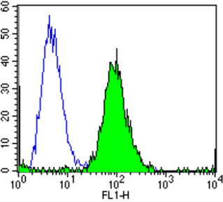
Flow cytometry analysis of Calmodulin showing positive staining in the cytoplasm of C6 cells compared to an isotype control (blue). Cells were harvested, adjusted to a concentration of 1-5x10^6 cells/ml, fixed with 2% paraformaldehyde and washed with PBS. Cells were penetrated by dropping the supernatant, adding 90% methanol and incubated for 10 minutes at room temperature. Follwing penetration, cells were blocked with a 2% solution of BSA-PBS for 30 min at room temperature and incubated with a Calmodulin monoclonal antibody at a dilution of 2 ug/test for 60 min at room temperature. Cells were then incubated for 40 min at room temperature in the dark using a Dylight 488-conjugated goat anti-mouse IgG (H+L) secondary antibody and re-suspended in PBS for FACS analysis.
Application
Immunofluorescent analysis of Calmodulin using Calmodulin Monoclonal antibody (2D1) shows staining in HeLa cells. Calmodulin staining (green), F-Actin staining with Phalloidin (red) and nuclei with DAPI (blue) is shown. Cells were grown on chamber slides and fixed with formaldehyde prior to staining. Cells were probed without (control) or with or an antibody recognizing Calmodulin at a dilution of 1:20 over night at 4 degrees C, washed with PBS and incubated with a DyLight-488 conjugated secondary antibody.
Application
Immunohistochemistry was performed on normal biopsies of deparaffinized Rat testis tissue. To expose target proteins, heat induced antigen retrieval was performed using 10mM sodium citrate (pH6.0) buffer, microwaved for 8-15 minutes. Following antigen retrieval tissues were blocked in 3% BSA-PBS for 30 minutes at room temperature. Tissues were then probed at a dilution of 1:20 with a mouse monoclonal antibody recognizing Calmodulin or without primary antibody (negative control) overnight at 4°C in a humidified chamber. Tissues were washed extensively with PBST and endogenous peroxidase activity was quenched with a peroxidase suppressor. Detection was performed using a biotin-conjugated secondary antibody and SA-HRP, followed by colorimetric detection using DAB. Tissues were counterstained with hematoxylin and prepped for mounting.
Application
Immunofluorescent analysis of Calmodulin using Calmodulin Monoclonal antibody (2D1) shows staining in A2058 melanoma cells. Calmodulin staining (green), F-Actin staining with Phalloidin (red) and nuclei with DAPI (blue) is shown. Cells were grown on chamber slides and fixed with formaldehyde prior to staining. Cells were probed without (control) or with or an antibody recognizing Calmodulin at a dilution of 1:20 over night at 4 degrees C, washed with PBS and incubated with a DyLight-488 conjugated secondary antibody.
Application
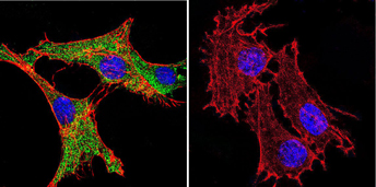
Immunofluorescent analysis of Calmodulin using Calmodulin Monoclonal antibody (2D1) shows staining in C6 glioma cells. Calmodulin staining (green), F-Actin staining with Phalloidin (red) and nuclei with DAPI (blue) is shown. Cells were grown on chamber slides and fixed with formaldehyde prior to staining. Cells were probed without (control) or with or an antibody recognizing Calmodulin at a dilution of 1:20 over night at 4 degrees C, washed with PBS and incubated with a DyLight-488 conjugated secondary antibody.
Application
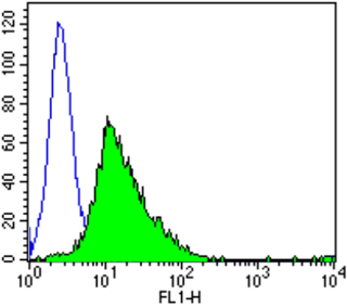
Flow cytometry analysis of Calmodulin showing positive staining in the cytoplasm of PC12 cells compared to an isotype control (blue). Cells were harvested, adjusted to a concentration of 1-5x10^6 cells/ml, fixed with 2% paraformaldehyde and washed with PBS. Cells were penetrated by dropping the supernatant, adding 90% methanol and incubated for 10 minutes at room temperature. Follwing penetration, cells were blocked with a 2% solution of BSA-PBS for 30 min at room temperature and incubated with a Calmodulin monoclonal antibody at a dilution of 2 ug/test for 60 min at room temperature. Cells were then incubated for 40 min at room temperature in the dark using a Dylight 488-conjugated goat anti-mouse IgG (H+L) secondary antibody and re-suspended in PBS for FACS analysis.
Application
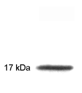
Western blot of purified calmodulin using
Application
Flow cytometry analysis of Calmodulin showing positive staining in the cytoplasm of MCF-7 cells compared to an isotype control (blue). Cells were harvested, adjusted to a concentration of 1-5x10^6 cells/ml, fixed with 2% paraformaldehyde and washed with PBS. Cells were penetrated by dropping the supernatant, adding 90% methanol and incubated for 10 minutes at room temperature. Follwing penetration, cells were blocked with a 2% solution of BSA-PBS for 30 min at room temperature and incubated with a Calmodulin monoclonal antibody at a dilution of 2 ug/test for 60 min at room temperature. Cells were then incubated for 40 min at room temperature in the dark using a Dylight 488-conjugated goat anti-mouse IgG (H+L) secondary antibody and re-suspended in PBS for FACS analysis.
Application
Immunohistochemistry was performed on normal biopsies of de-paraffinized Rat cerebellum tissue. To expose target proteins, heat induced antigen retrieval was performed using 10mM sodium citrate (pH6.0) buffer, microwaved for 8-15 minutes. Following antigen retrieval tissues were blocked in 3% BSA-PBS for 30 minutes at room temperature. Tissues were then probed at a dilution of 1:20 with a mouse monoclonal antibody recognizing Calmodulin or without primary antibody (negative control) overnight at 4°C in a humidified chamber. Tissues were washed extensively with PBST and endogenous peroxidase activity was quenched with a peroxidase suppressor. Detection was performed using a biotin-conjugated secondary antibody and SA-HRP, followed by colorimetric detection using DAB. Tissues were counterstained with hematoxylin and prepped for mounting.
Application
Immunolocalization of calmodulin in rat brain using| Application Notes |
For ICC/IF: Use at a dilution of 1:20 - 1:50. : |
|---|
| Form | Liquid |
|---|---|
| Storage Instructions | Keep as concentrated solution. Aliquot and store at -20ºC or below. Avoid multiple freeze-thaw cycles. |
| Storage Buffer | PBS containing 0.05% sodium azide |


