Application
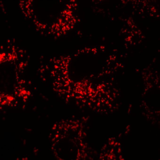
Immunocytochemical analysis of Calnexin in COS7 cells. Transfected COS7 cells were blocked in 1% BSA, 5% normal goat serum, and 0.1% Triton X-100 in 1X PBS, and then stained (1:500), followed by a fluorophore-conjugated secondary antibody (red). Magnification = 100X.Application
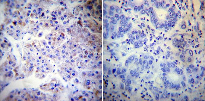
Immunohistochemistry was performed on cancer biopsies of deparaffinized human hepatocarcinoma tissues. To expose target proteins, heat induced antigen retrieval was performed using 10mM sodium citrate (pH6.0) buffer, microwaved for 8-15 minutes. Following antigen retrieval tissues were blocked in 3% BSA-PBS for 30 minutes at room temperature. Tissues were then probed at a dilution of 1:20 with or without Calnexin antibody [AF18] overnight at 4°C in a humidified chamber. Tissues were washed extensively with PBST and endogenous peroxidase activity was quenched with a peroxidase suppressor. Detection was performed using a biotin-conjugated secondary antibody and SA-HRP, followed by colorimetric detection using DAB. Tissues were counterstained with hematoxylin and prepped for mounting.| Product Name | Calnexin antibody |
|---|---|
| Antibody Type | Primary Antibodies |
| Product description | Mouse monoclonal antibody [AF18] to CalnexinThis gene encodes a member of the calnexin family of molecular chaperones. The encoded protein is a calcium-binding, endoplasmic reticulum (ER)-associated protein that interacts transiently with newly synthesized N-linked glycoproteins, facilitating protein folding and assembly. It may also play a central role in the quality control of protein folding by retaining incorrectly folded protein subunits within the ER for degradation. Alternatively spliced transcript variants encoding the same protein have been described. [provided by RefSeq, Jul 2008] |
| Immunogen | Focus human hepatoma cell lysate. |
| Clonality | Monoclonal |
|---|---|
| Isotype | IgG1 |
| Clone Number | AF18 |
| Host Species | Mouse |
| Tested Applications | ICC/IFIHCIHC-PIPWB |
| ICC/IF: 1:50 - 1:500: |
|
| Species Reactivity | HumanMouseRatPrimate |
| Concentration | 1mg/ml (Please refer to the vial label for the specific concentration) |
| Purification | Protein A |
| Gene Synonyms | P90 CNX IP90 CANX calnexin |
|---|---|
| Target | Calnexin |
Application
Immunocytochemical analysis of Calnexin in COS7 cells. Transfected COS7 cells were blocked in 1% BSA, 5% normal goat serum, and 0.1% Triton X-100 in 1X PBS, and then stained (1:500), followed by a fluorophore-conjugated secondary antibody (red). Magnification = 100X.
Application
Immunohistochemistry was performed on cancer biopsies of deparaffinized human hepatocarcinoma tissues. To expose target proteins, heat induced antigen retrieval was performed using 10mM sodium citrate (pH6.0) buffer, microwaved for 8-15 minutes. Following antigen retrieval tissues were blocked in 3% BSA-PBS for 30 minutes at room temperature. Tissues were then probed at a dilution of 1:20 with or without Calnexin antibody [AF18] overnight at 4°C in a humidified chamber. Tissues were washed extensively with PBST and endogenous peroxidase activity was quenched with a peroxidase suppressor. Detection was performed using a biotin-conjugated secondary antibody and SA-HRP, followed by colorimetric detection using DAB. Tissues were counterstained with hematoxylin and prepped for mounting.| Application Notes | ICC/IF: 1:50 - 1:500: |
|---|
| Form | Liquid |
|---|---|
| Storage Instructions | -20° C, Avoid Freeze/Thaw Cycles |
| Storage Buffer | PBS containing 0.05 % sodium azide |


