Application
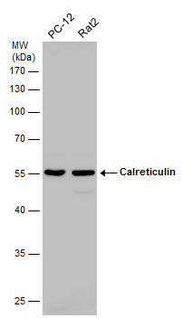
Various whole cell extracts (30 µg) were separated by 10% SDS-PAGE, and the membrane was blotted with Calreticulin antibody diluted at 1:1000. The HRP-conjugated anti-rabbit IgG antibody was used to detect the primary antibody.Application
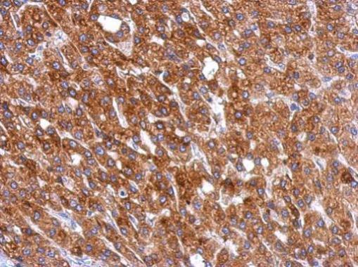
Immunohistochemical analysis of paraffin-embedded human hepatoma, using Calreticulin antibody at 1:500 dilution.Application
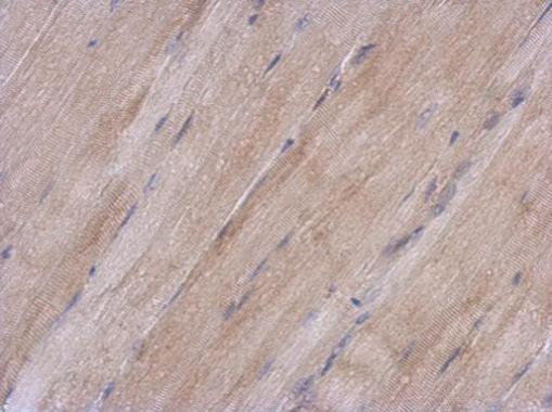
Immunohistochemical analysis of paraffin-embedded mouse muscle, using Calreticulin antibody at 1:500 dilution.Application
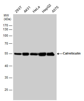
Calreticulin antibody detects Calreticulin protein by western blot analysis. Various whole cell extracts (30 µg) were separated by 10% SDS-PAGE, and the membrane was blotted with Calreticulin antibody diluted at a dilution of 1:1000. The HRP-conjugated anti-rabbit IgG antibody was used to detect the primary antibody.Application
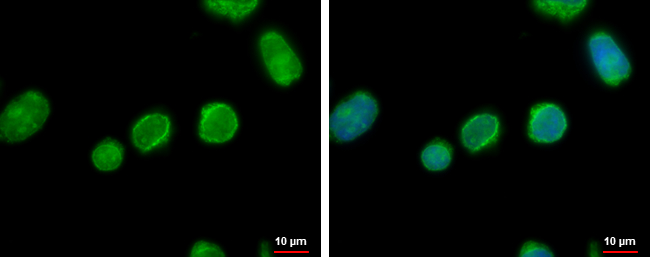
Calreticulin antibody detects Calreticulin protein at endoplasmic reticulum by immunofluorescent analysis. Sample: Jurkat cells were fixed in 4% paraformaldehyde at RT for 15 min. Green: Calreticulin protein stained by Calreticulin antibody diluted at 1:500. Blue: Hoechst 33342 staining. Scale bar = 10 μm.| Product Name | Calreticulin antibody |
|---|---|
| Antibody Type | Primary Antibodies |
| Product description | Rabbit Polyclonal antibody to CalreticulinCalreticulin is a multifunctional protein that acts as a major Ca(2+)-binding (storage) protein in the lumen of the endoplasmic reticulum. It is also found in the nucleus, suggesting that it may have a role in transcription regulation. Calreticulin binds to the synthetic peptide KLGFFKR, which is almost identical to an amino acid sequence in the DNA-binding domain of the superfamily of nuclear receptors. Calreticulin binds to antibodies in certain sera of systemic lupus and Sjogren patients which contain anti-Ro/SSA antibodies, it is highly conserved among species, and it is located in the endoplasmic and sarcoplasmic reticulum where it may bind calcium. The amino terminus of calreticulin interacts with the DNA-binding domain of the glucocorticoid receptor and prevents the receptor from binding to its specific glucocorticoid response element. Calreticulin can inhibit the binding of androgen receptor to its hormone-responsive DNA element and can inhibit androgen receptor and retinoic acid receptor transcriptional activities in vivo, as well as retinoic acid-induced neuronal differentiation. Thus, calreticulin can act as an important modulator of the regulation of gene transcription by nuclear hormone receptors. Systemic lupus erythematosus is associated with increased autoantibody titers against calreticulin but calreticulin is not a Ro/SS-A antigen. Earlier papers referred to calreticulin as an Ro/SS-A antigen but this was later disproven. Increased autoantibody titer against human calreticulin is found in infants with complete congenital heart block of both the IgG and IgM classes. [provided by RefSeq] |
| Immunogen | Recombinant protein encompassing a sequence within the center region of human Calreticulin. The exact sequence is proprietary. |
| Clonality | Polyclonal |
|---|---|
| Isotype | IgG |
| Host Species | Rabbit |
| Tested Applications | ICC/IFIHC-PWB |
| *Optimal dilutions/concentrations should be determined by the researcher.
: |
|
| Species Reactivity | HumanMouseRat |
| Predicted Reactivity | Zebrafish, Xenopus laevis, Pig, Rabbit, Rhesus Monkey, BovineZebrafish (81%), Xenopus laevis (83%), Pig (94%), Rabbit (95%), Rhesus Monkey (100%), Bovine (95%) |
| Concentration | 1mg/ml (Please refer to the vial label for the specific concentration) |
| Purification | Affinity purified |
| Gene Synonyms | CRT antibody FLJ26680 antibody RO antibody SSA antibody cC1qR antibody CALR antibody grp60 antibody endoplasmic reticulum resident protein 60 antibody calreticulin antibody calregulin antibody Sicca syndrome antigen A (autoantigen Ro calreticul |
|---|---|
| Molecular Weight(MW) | 48 kDa (note) |
| Cellular Localization | Endoplasmic reticulum lumen , Cytoplasm , cytosol , Secreted , extracellular space , extracellular matrix , Cell surface |
Application
Various whole cell extracts (30 µg) were separated by 10% SDS-PAGE, and the membrane was blotted with Calreticulin antibody diluted at 1:1000. The HRP-conjugated anti-rabbit IgG antibody was used to detect the primary antibody.
Application
Immunohistochemical analysis of paraffin-embedded human hepatoma, using Calreticulin antibody at 1:500 dilution.
Application
Immunohistochemical analysis of paraffin-embedded mouse muscle, using Calreticulin antibody at 1:500 dilution.
Application
Calreticulin antibody detects Calreticulin protein by western blot analysis. Various whole cell extracts (30 µg) were separated by 10% SDS-PAGE, and the membrane was blotted with Calreticulin antibody diluted at a dilution of 1:1000. The HRP-conjugated anti-rabbit IgG antibody was used to detect the primary antibody.
Application
Calreticulin antibody detects Calreticulin protein at endoplasmic reticulum by immunofluorescent analysis. Sample: Jurkat cells were fixed in 4% paraformaldehyde at RT for 15 min. Green: Calreticulin protein stained by Calreticulin antibody diluted at 1:500. Blue: Hoechst 33342 staining. Scale bar = 10 μm.| Positive Control | 293T , A431 , HeLa , HepG2 , A375 , Neuro 2A , C8D30 , NIH-3T3 , Raw264.7 , C2C12 , PC-12 , Rat2 |
|---|---|
| Application Notes | *Optimal dilutions/concentrations should be determined by the researcher.
: |
| Form | Liquid |
|---|---|
| Storage Instructions | Keep as concentrated solution. Aliquot and store at -20ºC or below. Avoid multiple freeze-thaw cycles. |
| Storage Buffer | 1XPBS, 20% Glycerol (pH7). 0.01% Thimerosal was added as a preservative. |


