Application
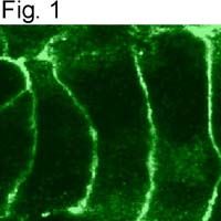
Immunofluorescent analysis of Cannabinoid Receptor II in AtT20 cells transfected with the rat CB2 gene.Application
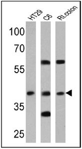
Western blot analysis of Cannabinoid Receptor II in 25 ug of HT29, C6 and rat colon lysates. Proteins were transferred to a PVDF membrane and blocked at 4°C overnight. The membrane was probed with Cannabinoid Receptor II antibody at a dilution of 1:200 overnight at 4°C, washed in TBST, and probed with an HRP-conjugated secondary antibody. Chemiluminescent detection was performed.Application
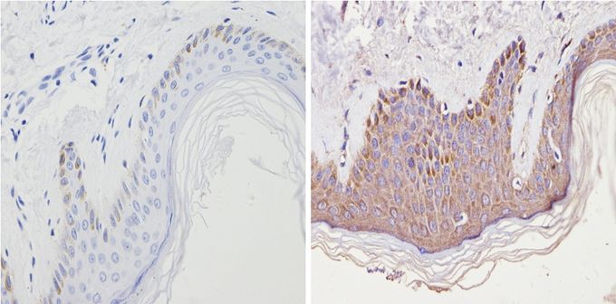
Immunohistochemistry analysis of Cannabinoid Receptor II in paraffin-embedded human skin tissue (right) compared with a negative control in the absence of primary antibody (left). To expose target proteins, antigen retrieval method was performed using 10mM sodium citrate (pH 6.0) microwaved for 8-15 min. Following antigen retrieval, tissues were blocked in 3% H2O2-methanol for 15 min at room temperature, washed with ddH2O and PBS, and then probed with Cannabinoid Receptor II antibody diluted by 3% BSA-PBS at a dilution of 1:20 overnight at 4°C in a humidified chamber. Tissues were washed extensively PBST and detection was performed using an HRP-conjugated secondary antibody followed by colorimetric detection using a DAB kit. Tissues were counterstained with hematoxylin and dehydrated with ethanol and xylene to prep for mounting.| Product Name | Cannabinoid Receptor 2 antibody |
|---|---|
| Antibody Type | Primary Antibodies |
| Product description | Rabbit polyclonal antibody to Cannabinoid Receptor 2G-protein-coupled transmembrane receptor; pertussis toxin-sensitive modulator of adenylate cyclase activity [RGD, Feb 2006] |
| Immunogen | A fusion protein containing the first 32 amino acid residues from rat Cannabinoid Receptor 2. |
| Clonality | Polyclonal |
|---|---|
| Isotype | IgG |
| Host Species | Rabbit |
| Tested Applications | FACSICC/IFIHCIHC-PWB |
| IHC-P: 1:10 - 1:100: |
|
| Species Reactivity | HumanMouseRat |
| Concentration | 1mg/ml |
| Purification | Affinity purified |
| Gene Synonyms | CB-2 CB2 cannabinoid receptor 2 (macrophage) Cnr2 CNR2C CB2C |
|---|---|
| Target | Cannabinoid Receptor 2 |
Application
Immunofluorescent analysis of Cannabinoid Receptor II in AtT20 cells transfected with the rat CB2 gene.
Application
Western blot analysis of Cannabinoid Receptor II in 25 ug of HT29, C6 and rat colon lysates. Proteins were transferred to a PVDF membrane and blocked at 4°C overnight. The membrane was probed with Cannabinoid Receptor II antibody at a dilution of 1:200 overnight at 4°C, washed in TBST, and probed with an HRP-conjugated secondary antibody. Chemiluminescent detection was performed.
Application
Immunohistochemistry analysis of Cannabinoid Receptor II in paraffin-embedded human skin tissue (right) compared with a negative control in the absence of primary antibody (left). To expose target proteins, antigen retrieval method was performed using 10mM sodium citrate (pH 6.0) microwaved for 8-15 min. Following antigen retrieval, tissues were blocked in 3% H2O2-methanol for 15 min at room temperature, washed with ddH2O and PBS, and then probed with Cannabinoid Receptor II antibody diluted by 3% BSA-PBS at a dilution of 1:20 overnight at 4°C in a humidified chamber. Tissues were washed extensively PBST and detection was performed using an HRP-conjugated secondary antibody followed by colorimetric detection using a DAB kit. Tissues were counterstained with hematoxylin and dehydrated with ethanol and xylene to prep for mounting.| Application Notes | IHC-P: 1:10 - 1:100: |
|---|
| Form | Liquid |
|---|---|
| Storage Instructions | -20° C, Avoid Freeze/Thaw Cycles |
| Storage Buffer | PBS containing 0.05 % sodium azide and 50 % glycerol |


