Application
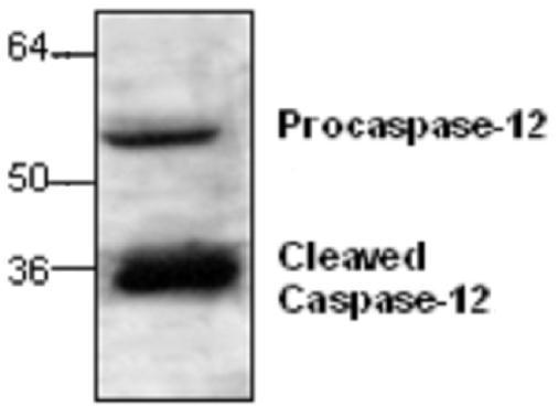
Western blot analysis of Caspase-12 with rat kidney tissue lysate.| Product Name | Caspase 12 antibody |
|---|---|
| Antibody Type | Primary Antibodies |
| Product description | Rabbit polyclonal antibody to Caspase 12Three distinct signaling pathways lead to programmed cell death (apoptosis). The death receptor and mitochondrion pathways are the mains two pathways, in which the key apoptotic proteases caspase-8 and caspase-9, respectively, are involved. The endoplasmic reticulum (ER) stress is the third apoptotic pathway and caspase-12 is involved. Caspase-12 is located to the ER, but not to the cytoplasm or mitochondrion. Caspase-12 is activated by ER stress, including disruption of ER calcium hemeostasis, and mediates ER stress-induced apoptosis. Caspase-12 is localized to the ER with several proteins that are involved in Alzheimer’s disease including gamma-secretase presenilin and beta-amyloid precursor protein (APP). Caspase-12 mediates cytotoxicity induced by amyloid-beta. Caspase-12 is ubiquitously expressed in mouse tissues. |
| Clonality | Polyclonal |
|---|---|
| Isotype | IgG |
| Host Species | Rabbit |
| Tested Applications | WB |
| Western blotting (0.5-4 µg/ml). It detects procaspase-12 (55 kDa) and cleavage caspase-12 (36 kDa) on SDS-PAGE immunoblots. However, the optimal conditions should be determined individually.: |
|
| Species Reactivity | MouseRat |
| Concentration | 0.2mg/ml |
| Purification | Affinity purified |
| Gene Synonyms | CASP12P1 CASP12 Caspase12 |
|---|---|
| Target | Caspase 12 |
| Tissue Specificity | This antibody is specific for Caspase-12. |
Application
Western blot analysis of Caspase-12 with rat kidney tissue lysate.| Application Notes | Western blotting (0.5-4 µg/ml). It detects procaspase-12 (55 kDa) and cleavage caspase-12 (36 kDa) on SDS-PAGE immunoblots. However, the optimal conditions should be determined individually.: |
|---|
| Form | Liquid |
|---|---|
| Storage Instructions | The antibody solution should be gently mixed before use. Stored at -20 °C for future use. |
| Storage Buffer | PBS (pH 7.2) containing 30% glycerol, 0.5% BSA, 0.01% thimerosal |


