Application
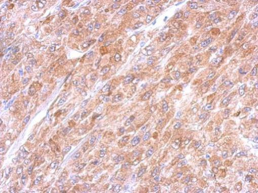
Immunohistochemical analysis of paraffin-embedded U87 xenograft, using Caspase 3 antibody at 1:500 dilution.Application
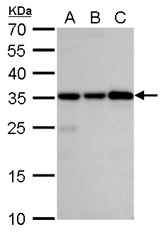
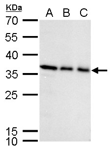
Caspase 3 antibody detects Caspase 3 protein by western blot analysis. A. 30 µg Jurkat whole cell lysate/extract B. 30 µg Raji whole cell lysate/extract C. 30 µg NCI-H929 whole cell lysate/extract 12% SDS-PAGE Caspase 3 antibody dilution: 1:2000 The HRP-conjugated anti-rabbit IgG antibody was used to detect the primary antibody.Application
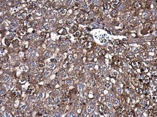
Caspase 3 antibody detects Caspase 3 protein at cytoplasm in rat liver by immunohistochemical analysis. Sample: Paraffin-embedded rat liver. Caspase 3 antibody diluted at 1:200.Application
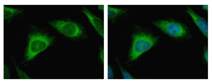
Caspase 3 antibody detects CASP3 protein at Cytoplasm by immunofluorescent analysis. Sample: HeLa cells were fixed in 4% paraformaldehyde at RT for 15 min. Green: CASP3 protein stained by Caspase 3 antibody diluted at 1:500. Blue: Hoechst 33343 staining.Application
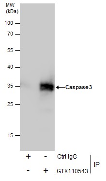
Immunoprecipitation of Caspase 3 protein from HeLa whole cell extracts using 5 μg of Caspase 3 antibody . Western blot analysis was performed using Caspase 3 antibody . EasyBlot anti-Rabbit IgG was used as a secondary reagent.Application
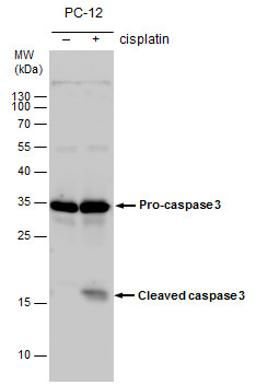
Caspase 3 antibody detects Caspase 3 protein by western blot analysis. Un-treated (-) and treated (+, 30 µM Cisplatin treatment for 24 hrs) PC-12 whole cell extracts (30 µg) were separated by 15% SDS-PAGE, and the membrane was blotted with Caspase 3 antibody diluted by 1:1000. The HRP-conjugated anti-rabbit IgG antibody was used to detect the primary antibody.Application
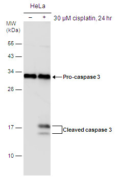
Untreated (–) and treated (+) HeLa whole cell extracts (30 µg) were separated by 12% SDS-PAGE, and the membrane was blotted with Caspase 3 antibody diluted at 1:1000. The HRP-conjugated anti-rabbit IgG antibody was used to detect the primary antibody.Application
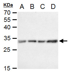
Caspase 3 antibody detects Caspase 3 protein by western blot analysis. A. 30 µg C8D30 whole cell extract B. 30 µg NIH-3T3 whole cell extract C. 30 µg Raw264.7 whole cell extract D. 30 µg C2C12 whole cell extract 12% SDS-PAGE Caspase 3 antibody dilution: 1:1000 The HRP-conjugated anti-rabbit IgG antibody was used to detect the primary antibody.Application
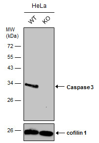
Wild-type (WT) and Caspase 3 knownout (KO) HeLa cell extracts (30 µg) were separated by 12% SDS-PAGE, and the membrane was blotted with Caspase 3 antibody diluted at 1:2000. The HRP-conjugated anti-rabbit IgG antibody was used to detect the primary antibody.Application
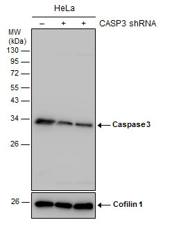
Non-transfected (–) and transfected (+) HeLa whole cell extracts (30 µg) were separated by 12% SDS-PAGE, and the membrane was blotted with Caspase 3 antibody diluted at 1:1000. The HRP-conjugated anti-rabbit IgG antibody was used to detect the primary antibody.Application
Caspase 3 antibody detects Caspase 3 protein by western blot analysis. A. 30 µg K562 whole cell lysate/extract B. 30 µg THP-1 whole cell lysate/extract C. 30 µg HL-60 whole cell lysate/extract 12% SDS-PAGE Caspase 3 antibody dilution: 1:1000 The HRP-conjugated anti-rabbit IgG antibody was used to detect the primary antibody.Application
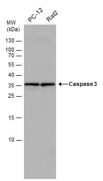
Caspase 3 antibody detects Caspase 3 protein by western blot analysis. Various whole cell extracts (30 µg) were separated by 15% SDS-PAGE, and the membrane was blotted with Caspase 3 antibody diluted by 1:1000. The HRP-conjugated anti-rabbit IgG antibody was used to detect the primary antibody.| Product Name | Caspase 3 antibody |
|---|---|
| Antibody Type | Primary Antibodies |
| Product description | Rabbit Polyclonal antibody to Caspase 3 (caspase 3, apoptosis-related cysteine peptidase)This gene encodes a protein which is a member of the cysteine-aspartic acid protease (caspase) family. Sequential activation of caspases plays a central role in the execution-phase of cell apoptosis. Caspases exist as inactive proenzymes which undergo proteolytic processing at conserved aspartic residues to produce two subunits, large and small, that dimerize to form the active enzyme. This protein cleaves and activates caspases 6, 7 and 9, and the protein itself is processed by caspases 8, 9 and 10. It is the predominant caspase involved in the cleavage of amyloid-beta 4A precursor protein, which is associated with neuronal death in Alzheimer's disease. Alternative splicing of this gene results in two transcript variants that encode the same protein. [provided by RefSeq] |
| Immunogen | Recombinant protein encompassing a sequence within the center region of human Caspase 3. The exact sequence is proprietary. |
| Modification | Acetylated |
| Clonality | Polyclonal |
|---|---|
| Isotype | IgG |
| Host Species | Rabbit |
| Tested Applications | ICC/IFIHC-FIHC-PIPWB |
| WB:1:500~1:2000 IP:1:100~1:500 ICC/IF:1:100~1:500 IHC:1:50~1:200 |
|
| Species Reactivity | HumanMouseRat |
| Predicted Reactivity | Dog, Cat, Pig, Rabbit, Chimpanzee, BovineDog (89%), Cat (87%), Pig (88%), Rabbit (86%), Chimpanzee (99%), Bovine (84%) |
| Concentration | 1mg/ml (Please refer to the vial label for the specific concentration) |
| Purification | Affinity purified |
| Gene Synonyms | CPP32 antibody CPP32B antibody SCA-1 antibody CASP3 antibody caspase-3 antibody apopain antibody procaspase3 antibody CASP-3 antibody protein Yama antibody |
|---|---|
| Molecular Weight(MW) | 32 kDa (note) (2) |
| Cellular Localization | Cytoplasm |
Application
Immunohistochemical analysis of paraffin-embedded U87 xenograft, using Caspase 3 antibody at 1:500 dilution.
Application
Caspase 3 antibody detects Caspase 3 protein by western blot analysis. A. 30 µg Jurkat whole cell lysate/extract B. 30 µg Raji whole cell lysate/extract C. 30 µg NCI-H929 whole cell lysate/extract 12% SDS-PAGE Caspase 3 antibody dilution: 1:2000 The HRP-conjugated anti-rabbit IgG antibody was used to detect the primary antibody.
Application
Caspase 3 antibody detects Caspase 3 protein at cytoplasm in rat liver by immunohistochemical analysis. Sample: Paraffin-embedded rat liver. Caspase 3 antibody diluted at 1:200.
Application
Caspase 3 antibody detects CASP3 protein at Cytoplasm by immunofluorescent analysis. Sample: HeLa cells were fixed in 4% paraformaldehyde at RT for 15 min. Green: CASP3 protein stained by Caspase 3 antibody diluted at 1:500. Blue: Hoechst 33343 staining.
Application
Immunoprecipitation of Caspase 3 protein from HeLa whole cell extracts using 5 μg of Caspase 3 antibody . Western blot analysis was performed using Caspase 3 antibody . EasyBlot anti-Rabbit IgG was used as a secondary reagent.
Application
Caspase 3 antibody detects Caspase 3 protein by western blot analysis. Un-treated (-) and treated (+, 30 µM Cisplatin treatment for 24 hrs) PC-12 whole cell extracts (30 µg) were separated by 15% SDS-PAGE, and the membrane was blotted with Caspase 3 antibody diluted by 1:1000. The HRP-conjugated anti-rabbit IgG antibody was used to detect the primary antibody.
Application
Untreated (–) and treated (+) HeLa whole cell extracts (30 µg) were separated by 12% SDS-PAGE, and the membrane was blotted with Caspase 3 antibody diluted at 1:1000. The HRP-conjugated anti-rabbit IgG antibody was used to detect the primary antibody.
Application
Caspase 3 antibody detects Caspase 3 protein by western blot analysis. A. 30 µg C8D30 whole cell extract B. 30 µg NIH-3T3 whole cell extract C. 30 µg Raw264.7 whole cell extract D. 30 µg C2C12 whole cell extract 12% SDS-PAGE Caspase 3 antibody dilution: 1:1000 The HRP-conjugated anti-rabbit IgG antibody was used to detect the primary antibody.
Application
Wild-type (WT) and Caspase 3 knownout (KO) HeLa cell extracts (30 µg) were separated by 12% SDS-PAGE, and the membrane was blotted with Caspase 3 antibody diluted at 1:2000. The HRP-conjugated anti-rabbit IgG antibody was used to detect the primary antibody.
Application
Non-transfected (–) and transfected (+) HeLa whole cell extracts (30 µg) were separated by 12% SDS-PAGE, and the membrane was blotted with Caspase 3 antibody diluted at 1:1000. The HRP-conjugated anti-rabbit IgG antibody was used to detect the primary antibody.
Application
Caspase 3 antibody detects Caspase 3 protein by western blot analysis. A. 30 µg K562 whole cell lysate/extract B. 30 µg THP-1 whole cell lysate/extract C. 30 µg HL-60 whole cell lysate/extract 12% SDS-PAGE Caspase 3 antibody dilution: 1:1000 The HRP-conjugated anti-rabbit IgG antibody was used to detect the primary antibody.
Application
Caspase 3 antibody detects Caspase 3 protein by western blot analysis. Various whole cell extracts (30 µg) were separated by 15% SDS-PAGE, and the membrane was blotted with Caspase 3 antibody diluted by 1:1000. The HRP-conjugated anti-rabbit IgG antibody was used to detect the primary antibody.| Positive Control | HeLa , C8D30 , NIH-3T3 , Raw264.7 , C2C12 , PC-12 , Rat2 , K562 , THP-1 , HL-60 , Jurkat , Raji , NCI-H929 , HeLa(30 µM cisplatin treatment for 24hr) , PC-12 (30uM cisplatin treatment for 24h) , *PA-1 , *C6 |
|---|---|
| Application Notes | WB:1:500~1:2000 IP:1:100~1:500 ICC/IF:1:100~1:500 IHC:1:50~1:200 |
| Form | Liquid |
|---|---|
| Storage Instructions | Keep as concentrated solution. Aliquot and store at -20ºC or below. Avoid multiple freeze-thaw cycles. |
| Storage Buffer | 1XPBS, 20% Glycerol (pH7). 0.025% ProClin 300 was added as a preservative. |


