Application
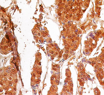
Formalin-fixed, paraffin-embedded human breast cancer stained with Caspase-3 antibody at 4 ug/ml. Localization can be cytoplasmic and nuclear. Staining in the nucleus is considered to be an indication of active Caspase-3. In most cell types and model systems, cells with active Caspase-3 are undergoing apoptosis. Cancer/normal adjacent tissue array was used for this test.Application
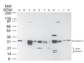
Western blot analysis of multiple human tissues using Caspase-3 antibody at 5 ug/ml. The tissues shown are A) brain, B) heart, C) intestine, D) kidney, E) liver, F) lung, G) muscle, H) stomach, I) spleen, J) ovary, and K) testis.Application
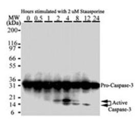
Western blot analysis of Caspase-3 in HeLa cells. Cells were treated with 2 uM staurosporine for different time periods. Caspase-3 activation is detected in Western blots by the presence of cleavage fragments. The antibody detected both pro (full-length) and active (cleaved) protein, depending on the treatment time points. Pro Caspase-3 is detected at ~32 kDa. Active/cleaved Caspase-3 (large subunit) is detected at ~14-21 kDa as one or more bands.Application
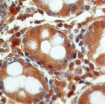
Formalin-fixed, paraffin-embedded normal colon stained with Caspase-3 antibody at 4 ug/ml. Localization can be cytoplasmic and nuclear. Staining in the nucleus is considered to be an indication of active Caspase-3. In most cell types and model systems, cells with active Caspase-3 are undergoing apoptosis. Cancer/normal adjacent tissue array was used for this test.| Product Name | Caspase 3 antibody |
|---|---|
| Antibody Type | Primary Antibodies |
| Product description | Mouse monoclonal antibody [31A1067] to Caspase 3Caspases are a family of cysteine proteases that are key mediators of programmed cell death or apoptosis. The precursor form of all caspases is composed of a prodomain, and large and small catalytic subunits. The active forms of caspases are generated by several stimuli including ligand-receptor interactions, growth factor deprivation and inhibitors of cellular functions. All known caspases require cleavage adjacent to aspartates to liberate one large and one small subunit, which associate into a2b2 tetramer to form the active enzyme. Gene for Caspase 3 also known as Yama, CPP32, and apopain codes for a 32-kDa protein. Caspase 3 cleaves the death substrate poly(ADP-ribose) polymerase (PARP) to a specific 85 kDa form observed during apoptosis and is inhibitable by the CrmA protein. Other Caspase 3 substrates include DNA-PK, actin, GAS2, and procaspase-6, etc. Caspase 3 is activated by cleavage events at Asp-28/Ser-29 (between N-terminal pro-domain) and Asp-175/Ser-176 (between large and small subunits) to generate a large subunit of 17-kDa and a small subunit of 12-kDa. |
| Immunogen | Recombinant fusion protein (6xHis tagged full-length human Caspase 3 protein expressed in bacteria). |
| Clonality | Monoclonal |
|---|---|
| Isotype | IgG1 |
| Clone Number | 31A1067 |
| Host Species | Mouse |
| Tested Applications | IHC-PWB |
| WB: Use at a concentration of 2 - 3 μg/ml. Predicted molecular weight: 31 kDa. Optimal dilutions/concentrations should be determined by the end user.: |
|
| Species Reactivity | Human |
| Concentration | 0.5mg/ml |
| Purification | Protein G |
| Gene Synonyms | 836 Caspase3 active & pro P42574 Caspase 3 active & pro CASP3 SCA1 CPP32B CPP32 SCA-1 600636 SCA 1 Caspase 3 (Active) Active Caspase 3 |
|---|---|
| Target | Caspase 3 |
| Cellular Localization | Cytoplasmic |
Application
Formalin-fixed, paraffin-embedded human breast cancer stained with Caspase-3 antibody at 4 ug/ml. Localization can be cytoplasmic and nuclear. Staining in the nucleus is considered to be an indication of active Caspase-3. In most cell types and model systems, cells with active Caspase-3 are undergoing apoptosis. Cancer/normal adjacent tissue array was used for this test.
Application
Western blot analysis of multiple human tissues using Caspase-3 antibody at 5 ug/ml. The tissues shown are A) brain, B) heart, C) intestine, D) kidney, E) liver, F) lung, G) muscle, H) stomach, I) spleen, J) ovary, and K) testis.
Application
Western blot analysis of Caspase-3 in HeLa cells. Cells were treated with 2 uM staurosporine for different time periods. Caspase-3 activation is detected in Western blots by the presence of cleavage fragments. The antibody detected both pro (full-length) and active (cleaved) protein, depending on the treatment time points. Pro Caspase-3 is detected at ~32 kDa. Active/cleaved Caspase-3 (large subunit) is detected at ~14-21 kDa as one or more bands.
Application
Formalin-fixed, paraffin-embedded normal colon stained with Caspase-3 antibody at 4 ug/ml. Localization can be cytoplasmic and nuclear. Staining in the nucleus is considered to be an indication of active Caspase-3. In most cell types and model systems, cells with active Caspase-3 are undergoing apoptosis. Cancer/normal adjacent tissue array was used for this test.| Positive Control | Staurosporine-treated HeLa or Jurkat cell lysate |
|---|---|
| Application Notes | WB: Use at a concentration of 2 - 3 μg/ml. Predicted molecular weight: 31 kDa. Optimal dilutions/concentrations should be determined by the end user.: |
| Form | Liquid |
|---|---|
| Storage Instructions | Keep as concentrated solution. Store at 4ºC short term. For extended storage aliquot and store at -20ºC or below. Avoid freeze-thaw cycles. |
| Storage Buffer | Preservative: 0.02% Sodium Azide; Constituents: PBS |


