Application
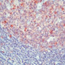
Formalin-fixed, paraffin-embedded human tonsil stained with using peroxidase-conjugate and AEC chromogen. Note cytoplasmic and nuclear staining of lymphoid cells.| Product Name | Caspase 3 antibody |
|---|---|
| Antibody Type | Primary Antibodies |
| Product description | Mouse monoclonal [3CSP01 ( 7.1.44 )] to Caspase 3Caspase 3 is ubiquitously expressed and like other caspases is synthesized as an inactive (32 kDa) proenzyme. Upon activation, Caspase 3 is cleaved at Asp 28 Ser 29 and Asp 175 Ser 176 thereby generating two subunits of 17kDa and 12kDa, respectively. Recent studies have implicated that Caspase 3 is associated with the induction of apoptosis. Activation of Caspase 3 occurs in response to variety of apoptotic inducers including Fas mediated apoptosis. |
| Immunogen | Recombinant full length protein (Human). |
| Clonality | Monoclonal |
|---|---|
| Isotype | IgG2a |
| Clone Number | 3CSP01 (7.1.44) |
| Host Species | Mouse |
| Tested Applications | IHC-PIPWB |
| Immunoprecipitation (native and denatured): use at a concentration of 2μg / mg of lysate. Western Blotting: use at a concentration of 1 - 2μg / ml for 2hrs at RT. Immunohistochemistry (formalin fixed paraffin embedded): use at a concentration of 2 - 4μg / ml for 30 min at RT. Optimal dilutions / concentrations should be determined by the end user. By Western blot, this antibody detects a band of 32 kDa, which corresponds to the predicted molecular weight of Caspase 3. Staining of formalin fixed tissues requires boiling tissue sections in 10mM citrate buffer, pH 6.0, for 10 - 20 min followed by cooling at RT for 20 min.: |
|
| Species Reactivity | Human |
| Concentration | 0.2mg/ml |
| Purification | Protein A |
| Gene Synonyms | 836 P42574 CASP3 Caspase3 SCA1 Caspase 3 CPP32B CPP32 SCA-1 600636 SCA 1 |
|---|---|
| Target | Caspase 3 |
| Cellular Localization | Predominantly Cytoplasmic with some nuclear staining. |
Application
Formalin-fixed, paraffin-embedded human tonsil stained with using peroxidase-conjugate and AEC chromogen. Note cytoplasmic and nuclear staining of lymphoid cells.| Positive Control | Jurkat cells. Tonsil and appendix tissue sections. |
|---|---|
| Application Notes | Immunoprecipitation (native and denatured): use at a concentration of 2μg / mg of lysate. Western Blotting: use at a concentration of 1 - 2μg / ml for 2hrs at RT. Immunohistochemistry (formalin fixed paraffin embedded): use at a concentration of 2 - 4μg / ml for 30 min at RT. Optimal dilutions / concentrations should be determined by the end user. By Western blot, this antibody detects a band of 32 kDa, which corresponds to the predicted molecular weight of Caspase 3. Staining of formalin fixed tissues requires boiling tissue sections in 10mM citrate buffer, pH 6.0, for 10 - 20 min followed by cooling at RT for 20 min.: |
| Form | Liquid |
|---|---|
| Storage Instructions | Keep as concentrated solution, aliquot and store at 4°C. Do not freeze. |
| Storage Buffer | 10mM PBS, pH7.4, 0.2%BSA, 0.09% sodium azide |


