Application
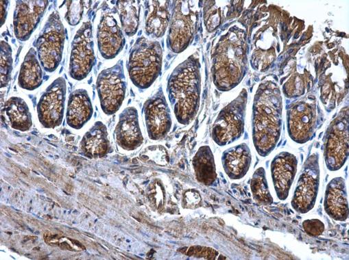
CD2AP antibody detects CD2AP protein at cytosol on mouse colon by immunohistochemical analysis. Sample: Paraffin-embedded mouse colon. CD2AP antibody dilution: 1:500.Application
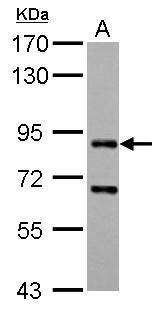
CD2AP antibody detects CD2AP protein by western blot analysis. A. 30 µg Rat2 whole cell lysate/extract 7.5% SDS-PAGE CD2AP antibody dilution: 1:1000 The HRP-conjugated anti-rabbit IgG antibody was used to detect the primary antibody.Application
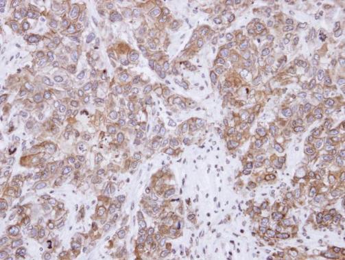
Immunohistochemical analysis of paraffin-embedded FaDu xenograft, using CD2-associated protein antibody at 1:500 dilution.Application
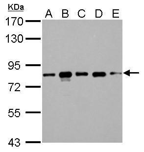
CD2AP antibody detects CD2AP protein by western blot analysis. A. 30 µg 293T whole cell lysate/extract B. 30 µg A431 whole cell lysate/extract C. 30 µg HeLa whole cell lysate/extract D. 30 µg HepG2 whole cell lysate/extract E. 30 µg A375 whole cell lysate/extract 7.5% SDS-PAGE CD2AP antibody dilution: 1:1000 The HRP-conjugated anti-rabbit IgG antibody was used to detect the primary antibody.Application
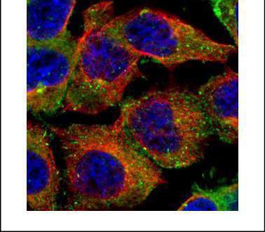
Confocal immunofluorescence analysis (Olympus FV10i) of paraformaldehyde-fixed A431, using CD2AP antibody (Green) at 1:500 dilution. Alpha-tubulin filaments were labeled with (Red) at 1:500.| Product Name | CD2AP antibody |
|---|---|
| Antibody Type | Primary Antibodies |
| Product description | Rabbit Polyclonal antibody to CD2AP (CD2-associated protein)This gene encodes a scaffolding molecule that regulates the actin cytoskeleton. The protein directly interacts with filamentous actin and a variety of cell membrane proteins through multiple actin binding sites, SH3 domains, and a proline-rich region containing binding sites for SH3 domains. The cytoplasmic protein localizes to membrane ruffles, lipid rafts, and the leading edges of cells. It is implicated in dynamic actin remodeling and membrane trafficking that occurs during receptor endocytosis and cytokinesis. Haploinsufficiency of this gene is implicated in susceptibility to glomerular disease. [provided by RefSeq] |
| Immunogen | Recombinant protein encompassing a sequence within the center region of human CD2AP. The exact sequence is proprietary. |
| Clonality | Polyclonal |
|---|---|
| Isotype | IgG |
| Host Species | Rabbit |
| Tested Applications | ICC/IFIHC-PWB |
| *Optimal dilutions/concentrations should be determined by the researcher.
: |
|
| Species Reactivity | HumanMouseRat |
| Predicted Reactivity | Pig, Rhesus Monkey, BovinePig (92%), Rhesus Monkey (96%), Bovine (90%) |
| Concentration | 1mg/ml (Please refer to the vial label for the specific concentration) |
| Purification | Affinity purified |
| Gene Synonyms | CMS antibody DKFZp586H0519 antibody CD2AP antibody CD2-associated protein antibody Cas ligand with multiple Src homology 3 (SH3) domains antibody cas ligand with multiple SH3 domains antibody adapter protein CMS antibody |
|---|---|
| Molecular Weight(MW) | 71 kDa (note) |
| Cellular Localization | Cytoplasm , cytoskeleton , Cell projection , ruffle |
Application
CD2AP antibody detects CD2AP protein at cytosol on mouse colon by immunohistochemical analysis. Sample: Paraffin-embedded mouse colon. CD2AP antibody dilution: 1:500.
Application
CD2AP antibody detects CD2AP protein by western blot analysis. A. 30 µg Rat2 whole cell lysate/extract 7.5% SDS-PAGE CD2AP antibody dilution: 1:1000 The HRP-conjugated anti-rabbit IgG antibody was used to detect the primary antibody.
Application
Immunohistochemical analysis of paraffin-embedded FaDu xenograft, using CD2-associated protein antibody at 1:500 dilution.
Application
CD2AP antibody detects CD2AP protein by western blot analysis. A. 30 µg 293T whole cell lysate/extract B. 30 µg A431 whole cell lysate/extract C. 30 µg HeLa whole cell lysate/extract D. 30 µg HepG2 whole cell lysate/extract E. 30 µg A375 whole cell lysate/extract 7.5% SDS-PAGE CD2AP antibody dilution: 1:1000 The HRP-conjugated anti-rabbit IgG antibody was used to detect the primary antibody.
Application
Confocal immunofluorescence analysis (Olympus FV10i) of paraformaldehyde-fixed A431, using CD2AP antibody (Green) at 1:500 dilution. Alpha-tubulin filaments were labeled with (Red) at 1:500.| Positive Control | 293T , A431 , HeLa , HepG2 , A375 , Rat2 |
|---|---|
| Application Notes | *Optimal dilutions/concentrations should be determined by the researcher.
: |
| Form | Liquid |
|---|---|
| Storage Instructions | Keep as concentrated solution. Aliquot and store at -20ºC or below. Avoid multiple freeze-thaw cycles. |
| Storage Buffer | 1XPBS, 1% BSA, 20% Glycerol (pH7). 0.01% Thimerosal was added as a preservative. |


