Application
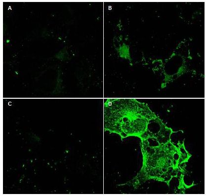
Immunofluorescence Microscopy using GeneTex Protein A purified anti-CD97 antibody shows staining of Fc-CD97-(5EGF) (panel D) in transfected COS cells. Panel A and C shows similar staining using pre-immune serum. Panel A and B show staining of mock transfected COS cells (no vector). A 1:2,500 dilution of the primary antibody was used.Application
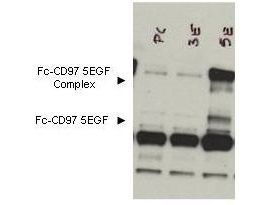
Western blot using GeneTex's Protein A purified anti-CD97 antibody shows detection of bands corresponding to free Fc-CD97- (5EGF) (lower arrowhead) and Fc-CD97- (5EGF) present as a complex (upper arrowhead) in lysates from COS cells. The left lane contains lysate from cells transfected with control DNA. The right lane contains lysate from COS cells expressing Fc-CD97- (5EGF). Approximately 10 ul of lysate was used in each lane. A 1:1,000 dilution of the primary antibody was used. The image was processed using a 10-sec exposure.| Product Name | Cd97 antibody |
|---|---|
| Antibody Type | Primary Antibodies |
| Product description | Rabbit Polyclonal antibody to Cd97Receptor potentially involved in both adhesion and signaling processes early after leukocyte activation. Plays an essential role in leukocyte migration. |
| Immunogen | This affinity purified antibody was prepared from whole rabbit serum produced by repeated immunizations with a recombinant protein corresponding to amino acids 1-512 (extracellular domain) of mouse CD97 protein. |
| Clonality | Polyclonal |
|---|---|
| Isotype | IgG |
| Host Species | Rabbit |
| Tested Applications | ELISAICC/IFWB |
| *Optimal dilutions/concentrations should be determined by the researcher.
: |
|
| Species Reactivity | HumanChimpanzee |
| Concentration | 1mg/ml |
| Purification | Protein A |
| Gene Synonyms | AA409984 TM7LN1 ADGRE5 |
|---|---|
| Target | Cd97 |
Application
Immunofluorescence Microscopy using GeneTex Protein A purified anti-CD97 antibody shows staining of Fc-CD97-(5EGF) (panel D) in transfected COS cells. Panel A and C shows similar staining using pre-immune serum. Panel A and B show staining of mock transfected COS cells (no vector). A 1:2,500 dilution of the primary antibody was used.
Application
Western blot using GeneTex's Protein A purified anti-CD97 antibody shows detection of bands corresponding to free Fc-CD97- (5EGF) (lower arrowhead) and Fc-CD97- (5EGF) present as a complex (upper arrowhead) in lysates from COS cells. The left lane contains lysate from cells transfected with control DNA. The right lane contains lysate from COS cells expressing Fc-CD97- (5EGF). Approximately 10 ul of lysate was used in each lane. A 1:1,000 dilution of the primary antibody was used. The image was processed using a 10-sec exposure.| Application Notes | *Optimal dilutions/concentrations should be determined by the researcher.
: |
|---|
| Form | Liquid |
|---|---|
| Storage Instructions | Store vial at 4° C for short-term use. For extended storage aliquot contents and freeze at -20° C or below. Avoid cycles of freezing and thawing. |
| Storage Buffer | 0.02 M Potassium Phosphate, 0.15 M Sodium Chloride, pH 7.2 and 0.01% (w/v) Sodium Azide |


