Application
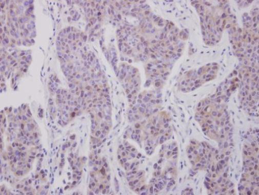
Immunohistochemical analysis of paraffin-embedded human breast cancer, using CDK7 antibody at 1:250 dilution.Application
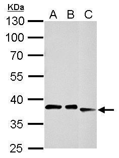
CDK7 antibody detects CDK7 protein by western blot analysis. A. 30 µg 293T whole cell lysate/extract B. 30 µg A431 whole cell lysate/extract C. 30 µg A375 whole cell lysate/extract 10% SDS-PAGE CDK7 antibody dilution: 1:1000 The HRP-conjugated anti-rabbit IgG antibody was used to detect the primary antibody.Application
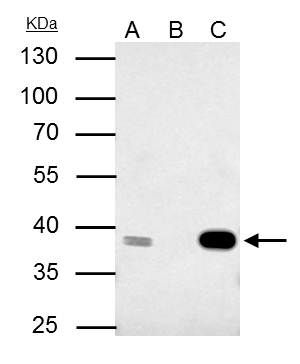
CDK7 antibody immunoprecipitates CDK7 protein in IP experiments. IP samples: 293T whole cell extract A. 40 μg 293T whole cell extract B. Control with 4 μg of preimmune Rabbit IgG C. Immunoprecipitation of CDK7 protein by 4 μg CDK7 antibody 10 % SDS-PAGE The immunoprecipitated CDK7 protein was detected by CDK7 antibody diluted at 1:500. [EasyBlot anti-rabbit IgG was used as a secondary reagent]Application
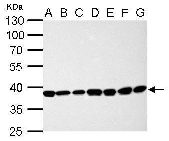
CDK7 antibody detects CDK7 protein by western blot analysis. A. 30 µg Neuro2A whole cell lysate/extract B. 30 µg GL261 whole cell lysate/extract C. 30 µg C8D30 whole cell lysate/extract D. 30 µg NIH-3T3 whole cell lysate/extract E. 30 µg BCL-1 whole cell lysate/extract F. 30 µg Raw264.7 whole cell lysate/extract G. 30 µg C2C12 whole cell lysate/extract 10% SDS-PAGE CDK7 antibody dilution: 1:1000 The HRP-conjugated anti-rabbit IgG antibody was used to detect the primary antibody.| Product Name | CDK7 antibody |
|---|---|
| Antibody Type | Primary Antibodies |
| Product description | Rabbit Polyclonal antibody to CDK7 (cyclin-dependent kinase 7)The protein encoded by this gene is a member of the cyclin-dependent protein kinase (CDK) family. CDK family members are highly similar to the gene products of Saccharomyces cerevisiae cdc28, and Schizosaccharomyces pombe cdc2, and are known to be important regulators of cell cycle progression. This protein forms a trimeric complex with cyclin H and MAT1, which functions as a Cdk-activating kinase (CAK). It is an essential component of the transcription factor TFIIH, that is involved in transcription initiation and DNA repair. This protein is thought to serve as a direct link between the regulation of transcription and the cell cycle. [provided by RefSeq] |
| Immunogen | Full length human CDK7 Recombinant protein. |
| Clonality | Polyclonal |
|---|---|
| Isotype | IgG |
| Host Species | Rabbit |
| Tested Applications | IHC-PIPWB |
| *Optimal dilutions/concentrations should be determined by the researcher.
: |
|
| Species Reactivity | HumanMouse |
| Predicted Reactivity | Zebrafish, Xenopus laevis, Pig, Rhesus Monkey, BovineZebrafish (85%), Xenopus laevis (86%), Pig (97%), Rhesus Monkey (99%), Bovine (96%) |
| Concentration | 1mg/ml (Please refer to the vial label for the specific concentration) |
| Purification | Affinity purified |
| Gene Synonyms | CAK1 antibody CDKN7 antibody MO15 antibody STK1 antibody p39MO15 antibody CDK7 antibody TFIIH basal transcription factor complex kinase subunit antibody cell division protein kinase 7 antibody homolog of Xenopus MO15 Cdk-activating kinase antibody |
|---|---|
| Molecular Weight(MW) | 39 kDa (note) |
| Cellular Localization | Nucleus |
Application
Immunohistochemical analysis of paraffin-embedded human breast cancer, using CDK7 antibody at 1:250 dilution.
Application
CDK7 antibody detects CDK7 protein by western blot analysis. A. 30 µg 293T whole cell lysate/extract B. 30 µg A431 whole cell lysate/extract C. 30 µg A375 whole cell lysate/extract 10% SDS-PAGE CDK7 antibody dilution: 1:1000 The HRP-conjugated anti-rabbit IgG antibody was used to detect the primary antibody.
Application
CDK7 antibody immunoprecipitates CDK7 protein in IP experiments. IP samples: 293T whole cell extract A. 40 μg 293T whole cell extract B. Control with 4 μg of preimmune Rabbit IgG C. Immunoprecipitation of CDK7 protein by 4 μg CDK7 antibody 10 % SDS-PAGE The immunoprecipitated CDK7 protein was detected by CDK7 antibody diluted at 1:500. [EasyBlot anti-rabbit IgG was used as a secondary reagent]
Application
CDK7 antibody detects CDK7 protein by western blot analysis. A. 30 µg Neuro2A whole cell lysate/extract B. 30 µg GL261 whole cell lysate/extract C. 30 µg C8D30 whole cell lysate/extract D. 30 µg NIH-3T3 whole cell lysate/extract E. 30 µg BCL-1 whole cell lysate/extract F. 30 µg Raw264.7 whole cell lysate/extract G. 30 µg C2C12 whole cell lysate/extract 10% SDS-PAGE CDK7 antibody dilution: 1:1000 The HRP-conjugated anti-rabbit IgG antibody was used to detect the primary antibody.| Positive Control | 293T , A431 , A375 , Neuro2A , GL261 , C8D30 , NIH-3T3 , BCL-1 , Raw264.7 , C2C12 |
|---|---|
| Application Notes | *Optimal dilutions/concentrations should be determined by the researcher.
: |
| Form | Liquid |
|---|---|
| Storage Instructions | Keep as concentrated solution. Aliquot and store at -20ºC or below. Avoid multiple freeze-thaw cycles. |
| Storage Buffer | 0.1M Tris, 0.1M Glycine, 10% Glycerol (pH7). 0.01% Thimerosal was added as a preservative. |


