Application
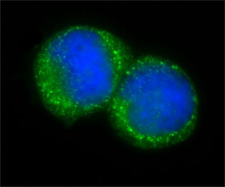
HeLa cells were fixed and stained with 6 μg/mL Anti-Centrin . The antibody was developed using Goat Anti-Rabbit IgG, Atto488 conjugate. Nuclear staining with DAPI.Application
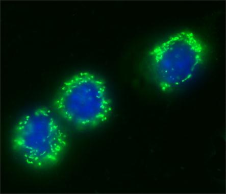
NIH3T3 cells were fixed and stained with 6 μg/mL Anti-Centrin . The antibody was developed using Goat Anti-Rabbit IgG, Atto488 conjugate. Nuclear staining with DAPI.Application
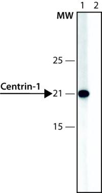
Lysate of HEK-293T cells overexpressing human centrin-1 was separated on SDS-PAGE, blotted with Anti-Centrin and developed using Goat Anti-Rabbit IgG-Peroxidase and a chemiluminescent substrate. Lanes 1. Antibody 0.3 μg/mL 2. Antibody 0.3 μg/mL + centrin1 immunizing peptide (human, 157-172)Application
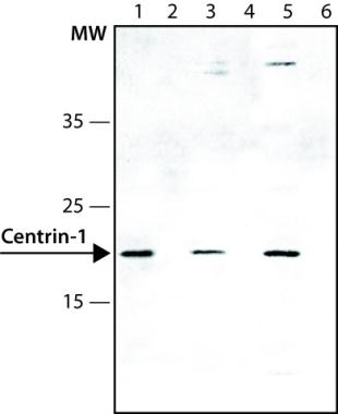
Lysates of HT-29 (lanes 1,2), HEK-293T (lanes (3-4) and HeLa (lanes 5-6) cells were separated on SDS-PAGE, blotted with 0.3 μg/mL Anti-Centrin and developed using Goat Anti-Rabbit IgG-Peroxidase and a chemiluminescent substrate. Lanes 1,3,5. Antibody 0.3 μg/mL 2,4,6. Antibody 0.3 μg/mL + centrin1 immunizing peptide (human, 157-172)| Product Name | Centrin 1 antibody |
|---|---|
| Antibody Type | Primary Antibodies |
| Product description | Rabbit polyclonal to Centrin 1This protein plays an important role in the determination of centrosome position and segregation, and in the process of microtubule severing. This encoded protein is localized to the centrosome of interphase cells, and redistributes to the region of the spindle poles during mitosis, reflecting the dynamic behavior of the centrosome during the cell cycle. |
| Immunogen | Synthetic peptide conjugated to KLH, corresponding to amino acids 157-172 of Human Centrin 1, with N-terminally added lysine. This sequence is highly conserved in centrin 1 of many species, e.g. mouse, drosophila, and Xenopus. It is also found in human an |
| Clonality | Polyclonal |
|---|---|
| Isotype | IgG |
| Host Species | Rabbit |
| Tested Applications | ICC/IFWB |
| WB: Use at a dilution of 1/2000, this concentration is recommended using a nuclear extract of the human epitheloid carcinoma HeLa cell line. Predicted molecular weight: 21 kDa. Optimal dilutions/concentrations should be determined by the end user.: |
|
| Species Reactivity | HumanMouseXenopus |
| Concentration | 1mg/ml |
| Purification | Affinity purified |
| Gene Synonyms | Centrin 1 CETN1 1068 Centrin1 Q12798 CEN1 CETN 603187 |
|---|---|
| Target | Centrin 1 |
| Cellular Localization | Centrosome of interphase and mitotic cells. |
Application
HeLa cells were fixed and stained with 6 μg/mL Anti-Centrin . The antibody was developed using Goat Anti-Rabbit IgG, Atto488 conjugate. Nuclear staining with DAPI.
Application
NIH3T3 cells were fixed and stained with 6 μg/mL Anti-Centrin . The antibody was developed using Goat Anti-Rabbit IgG, Atto488 conjugate. Nuclear staining with DAPI.
Application
Lysate of HEK-293T cells overexpressing human centrin-1 was separated on SDS-PAGE, blotted with Anti-Centrin and developed using Goat Anti-Rabbit IgG-Peroxidase and a chemiluminescent substrate. Lanes 1. Antibody 0.3 μg/mL 2. Antibody 0.3 μg/mL + centrin1 immunizing peptide (human, 157-172)
Application
Lysates of HT-29 (lanes 1,2), HEK-293T (lanes (3-4) and HeLa (lanes 5-6) cells were separated on SDS-PAGE, blotted with 0.3 μg/mL Anti-Centrin and developed using Goat Anti-Rabbit IgG-Peroxidase and a chemiluminescent substrate. Lanes 1,3,5. Antibody 0.3 μg/mL 2,4,6. Antibody 0.3 μg/mL + centrin1 immunizing peptide (human, 157-172)| Positive Control | HeLa cell line |
|---|---|
| Application Notes | WB: Use at a dilution of 1/2000, this concentration is recommended using a nuclear extract of the human epitheloid carcinoma HeLa cell line. Predicted molecular weight: 21 kDa. Optimal dilutions/concentrations should be determined by the end user.: |
| Form | Liquid |
|---|---|
| Storage Instructions | Keep as concentrated solution. Store at 4ºC short term. For extended storage aliquot and store at -20ºC or below. Avoid freeze-thaw cycles. |
| Storage Buffer | Preservative: 15mM Sodium Azide; Constituents: 1% BSA, 0.01M PBS. pH 7.4 |


