Application
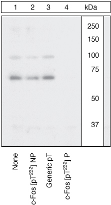
Extracts of SKBR3 cells were treated with 200ng/mL EGF for 10 minutes and resolved by SDS-PAGE on a 10% Tris-glycine gel and transferred to PVDF. The membrane was blocked with a 3% Milk-TBST buffer for one hour at room temperature, then incubated with the c-Fos [pT232] antibody for two hours at room temperature in a 3% Milk-TBST buffer, following prior incubation with: no peptide (1), the non-phosphopeptide corresponding to the phosphopeptide immunogen (2), a generic phosphothreonine-containing peptide (3), or the phosphopeptide immunogen (4). After washing, the membrane was incubated with goat F(ab)2 anti-rabbit IgG HRP conjugate and signals were detected. The data show that only the phosphopeptide corresponding to c-Fos [pT232] blocks the signal, demonstrating the specificity of the antibody. While c-Fos [pT232] is phosphorylated in the basal state of SKBR3 (data not shown), EGF treatment induced an increase in the phosphorylation signal.| Product Name | c-Fos (phospho Thr232) antibody |
|---|---|
| Antibody Type | Primary Antibodies |
| Product description | Rabbit polyclonal to c-Fos (phospho T232)The Fos gene family consists of 4 members: Fos, FosB, FosL1, and FosL2. These genes encode leucine zipper proteins that can dimerize with proteins of the JUN family, thereby forming the transcription factor complex AP1. As such, the Fos proteins have been implicated as regulators of cell proliferation, differentiation, and transformation. In some cases, expression of the Fos gene has also been associated with apoptotic cell death. The human oncogene c-Fos is homologous to the Finkel-Biskis-Jinkins (FBJ) murine osteosarcoma virus oncogene. Fos was the first transcription factor identified that has a critical function in regulating the development of cells destined to form and maintain the skeleton. |
| Immunogen | Synthetic peptide derived from the region of human c-Fos that contains Threonine 232. The sequence is conserved in mouse and rat. |
| Modification | p-Thr232 |
| Clonality | Polyclonal |
|---|---|
| Isotype | IgG |
| Host Species | Rabbit |
| Tested Applications | ICC/IFWB |
| WB: 1/1000. Predicted molecular weight: 41 kDa. Optimal dilutions/concentrations should be determined by the end user.: |
|
| Species Reactivity | HumanMouseRat |
| Concentration | 1mg/ml (Please refer to the vial label for the specific concentration) |
| Purification | Affinity purified |
| Gene Synonyms | 2353 FOS 164810 c-fos cFos P01100 c Fos c-Fos (phospho T232) c-Fos (phospho Thr232) c-Fos (pT232) c-Fos (pThr232) c-Fos phospho T232 c-Fos phospho Thr232 Phospho c-Fos (T232) Phospho c-Fos (Thr232) Phospho c-Fos (pT232) Phospho c-Fos (pThr |
|---|---|
| Target | c-Fos |
| Cellular Localization | Nuclear |
Application
Extracts of SKBR3 cells were treated with 200ng/mL EGF for 10 minutes and resolved by SDS-PAGE on a 10% Tris-glycine gel and transferred to PVDF. The membrane was blocked with a 3% Milk-TBST buffer for one hour at room temperature, then incubated with the c-Fos [pT232] antibody for two hours at room temperature in a 3% Milk-TBST buffer, following prior incubation with: no peptide (1), the non-phosphopeptide corresponding to the phosphopeptide immunogen (2), a generic phosphothreonine-containing peptide (3), or the phosphopeptide immunogen (4). After washing, the membrane was incubated with goat F(ab)2 anti-rabbit IgG HRP conjugate and signals were detected. The data show that only the phosphopeptide corresponding to c-Fos [pT232] blocks the signal, demonstrating the specificity of the antibody. While c-Fos [pT232] is phosphorylated in the basal state of SKBR3 (data not shown), EGF treatment induced an increase in the phosphorylation signal.| Positive Control | SKBR3 cells treated with EGF |
|---|---|
| Application Notes | WB: 1/1000. Predicted molecular weight: 41 kDa. Optimal dilutions/concentrations should be determined by the end user.: |
| Form | Liquid |
|---|---|
| Storage Instructions | Keep as concentrated solution. Store at 4ºC short term. For extended storage aliquot and store at -20ºC or below. Avoid freeze-thaw cycles. |
| Storage Buffer | PBS pH 7.3 (without Mg2+ and Ca2+) containing 50% glycerol, 1.0mg/ml BSA (IgG, protease free). and 0.05% sodium azide. |


